
Fig. 5
- ID
- ZDB-IMAGE-130910-5
- Publication
- Huang et al., 2013 - Treatment of Glucocorticoids Inhibited Early Immune Responses and Impaired Cardiac Repair in Adult Zebrafish
- All Figures
- Figures for Huang et al., 2013
|
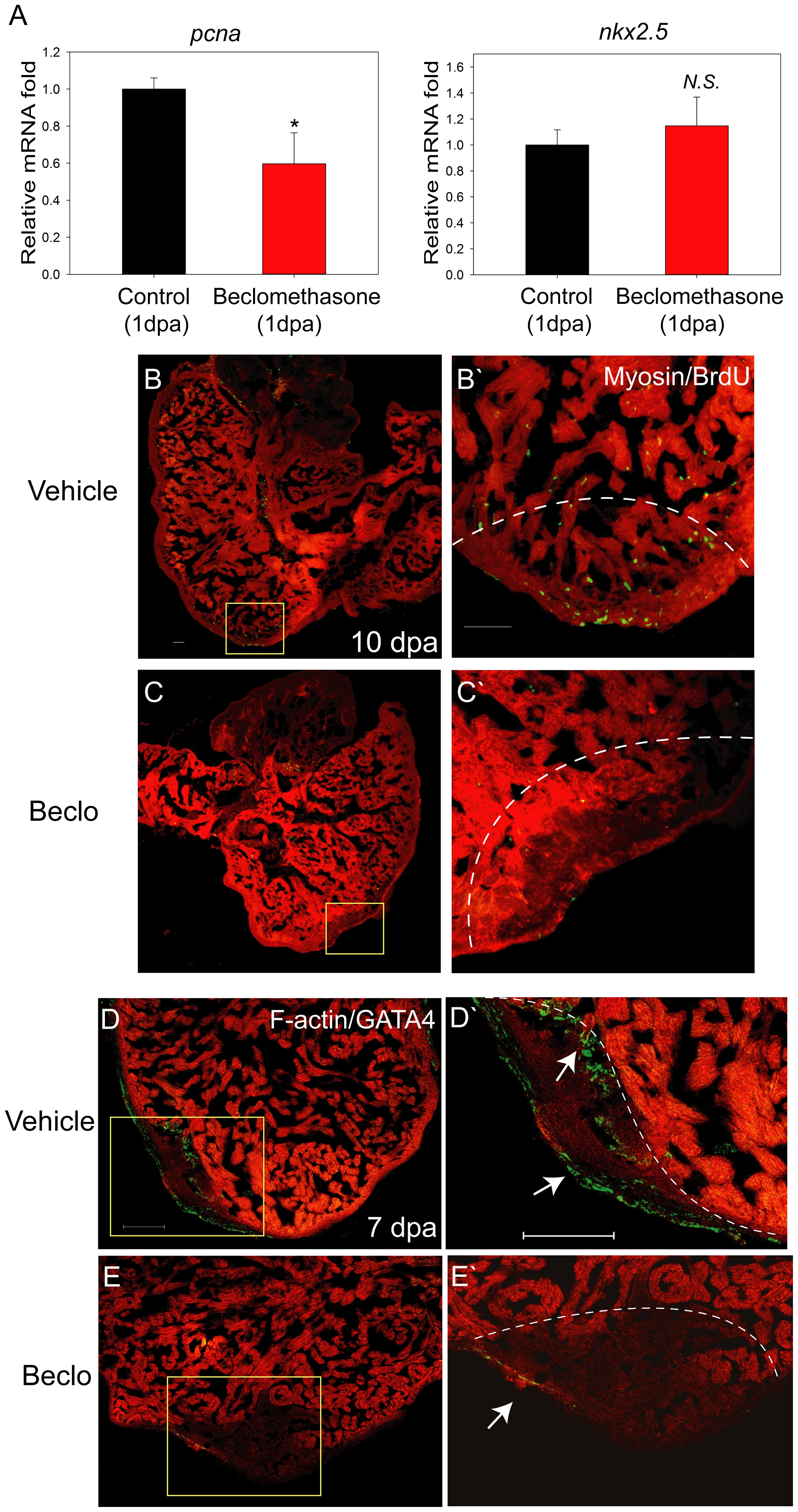
Fig. 5
Cell proliferation was hindered in the beclomethasone-treated zebrafish during cardiac repair.
Cell proliferation was examined in early phase of regeneration. (A) At 1 dpa, mRNA expression of proliferation marker gene, pcna, was reduced after beclomethasone treatment (40.3% reduction). In contract, the expression of the pre-cardiac gene, nkx2.5, showed no significant difference between the two groups. The data represent the mean±SEM. *indicates p<0.05. N.S. indicates no significant difference. (B, B′) In the control animals, numerous proliferating BrdU+ cells could be detected in the wound (n = 6). The dashed lines indicate the approximate amputation plane. (C, C′) The beclomethasone-treated animals showed little cell proliferation activities (n = 7, scale bar = 50 μm). (D, D′) In control groups, GATA4+ regenerating cells appeared near the injury site at 7 dpa. (arrow, n = 5) (E, E′) Beclomethasone treatment significantly reduced the GATA4+ regenerating cells during cardiac repair (arrow, n = 5, scale bar = 100 μm). The dashed lines indicate the approximate amputation plane.