
- Title
-
Integration of Microfractionation, qNMR and Zebrafish Screening for the In Vivo Bioassay-Guided Isolation and Quantitative Bioactivity Analysis of Natural Products
- Authors
- Bohni, N., Cordero-Maldonado, M.L., Maes, J., Siverio-Mota, D., Marcourt, L., Munck, S., Kamuhabwa, A.R., Moshi, M.J., Esguerra, C.V., de Witte, P.A., Crawford, A.D., and Wolfender, J.L.
- Source
- Full text @ PLoS One
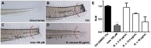
Anti-inflammatory activity of the methanolic extract of Rhynchosia viscosa. Anti-inflammatory activity was determined in an acute inflammation assay based on tail transection and treatment with lipopolysaccharides (LPS). A to D, zebrafish larvae are 4 days post-fertilization (dpf) with anterior to the left, scale bar = 10 μm. After tail transection and LPS exposure, stained leukocytes appear as black-brown spots migrating to the injured area in the transected tails. Migrating leukocytes were counted on one side in the tail in the region to the right of the dashed red arc and migration values were expressed as relative leukocyte migration (RLM) (E). A, tail of an uncut larva; B, negative control (DMSO 1%); C, positive control (indomethacin 100 μM) D, crude extract of R. viscosa at 50 μg/mL; E, graph displaying the RLM of 4 dpf larvae (n = 10) subjected to tail transection and incubation with R. viscosa. RLM d0.5 was established as cutoff for anti-inflammatory activity. * p<0.05. |

Anti-angiogenic activity of the methanolic extract of Rhynchosia viscosa. Inhibition of vascular outgrowth was determined in fli-1:EGFP transgenic embryos. At 16 hours post-fertilization (hpf), embryos were incubated with different concentrations of the methanolic extract of the plant and anti-angiogenic effects were assessed at 48 hpf. A to C, all embryos are 48 hpf, with anterior to the left, scale bar = 10 μm. A, untreated control (DMSO 1%); B, zoom of A (dashed box) showing normal outgrowth of intersegmental vessels (ISV) along the trunk of the larva (arrows); C, embryo treated with 50 μg/mL crude methanolic extract of R. viscosa. Inhibition or reduction of ISV growth is observed along the trunk (arrows); D, IC50 curve and values showing the inhibitory activity of the methanolic extract of R. viscosa. |

Bioactive compounds of Rhynchosia viscosa in the vascular outgrowth assay. IC50 curves and values were determined for each of the bioactive constituents of the methanolic extract of R. viscosa. Each compound, at six different concentrations, was assessed for their effect in the inhibition of intersegmental vessel (ISV) growth. A to F, all embryos are 48 hours post-fertilization (hpf), with anterior to the left, scale bar = 10 μm. A, untreated control (DMSO 1%); B, zoom of A (dashed box) showing normal outgrowth of intersegmental vessels (ISV) along the trunk of the larva (arrows); C, embryo treated with 50 μM genistein; D, embryo treated with 100 μM rhynchoviscin; E, embryo treated with 50 μM licoisoflavone A; F, embryo treated with 50 μM sophoraisoflavone. Arrowheads point the interconnection zone between the dorsal aorta and the posterior cardinal vein, arrows point the ISV; G, IC50 curves and values (μM) for each of the bioactive compounds of R. viscosa. |
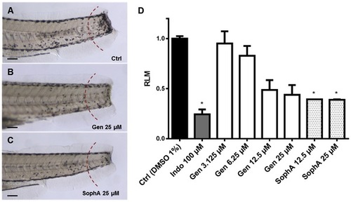
Anti-inflammatory effect of genistein and sophoraisoflavone A. A to C, zebrafish larvae are 4 dpf (days post-fertilization) with anterior to the left, scale bar = 10 μm. Migrating leukocytes were counted on one side in the tail in the region to the right of the dashed red arc and migration values were expressed as relative leukocyte migration (RLM) (C). A, negative control (DMSO 1%); B, genistein 25 μM; C, sophoraisoflavone A 25 μM; D, graph displaying the RLM in 4 dpf larvae (n = 10) after treatment with genistein and sophoraisoflavone A. RLM d0.5 was established as cutoff for anti-inflammatory activity. * p<0.05. |