
- Title
-
Knock-down of Cathepsin D in zebrafish fertilized eggs determines congenital myopathy
- Authors
- Follo, C., Ozzano, M., Montalenti, C., Santoro, M.M., and Isidoro, C.
- Source
- Full text @ Biosci. Rep.
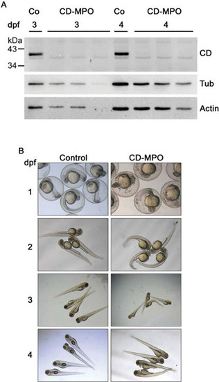
CD-knockdown zebrafish morphants exhibit a bent phenotype (A) Fertilized eggs at the stage of one or two cells were injected with CD–MPO or control MPO (Co). A pool of (not de-yolked) larvae at 3 and 4 dpf was collected and analysed by Western blot for CD expression. The Western blot of three different pools of larvae from three different experiments is shown. In control-injected larvae, the 41 kDa mature CD was detected, whereas in CD–MPO-injected larvae CD was absent. Tubulin and actin were used as reference of protein loading in the lanes. (B) Control and CD–MPO morphants at 1–4 dpf were imaged under the stereo microscope. A representative selection from three different experiments is shown. The bent phenotype is clearly evident in CD–MPO morphants. |
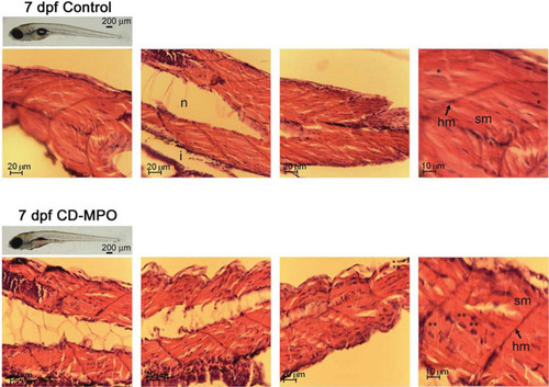
Histochemistry of the musculature in 7 dpf control and CD–MPO zebrafish Control and CD–MPO-injected zebrafish at 7 dpf showing an apparent ‘quasi-normal’ phenotype were subjected to longitudinal sectioning and H&E staining of the musculature. A selection of images from three independent experiments is shown. Symbols: hm, horizontal myoseptum; i, intestine; n, notochord; sm, somitic muscle.*syncytial nucleus; **eccentric nucleus. PHENOTYPE:
|
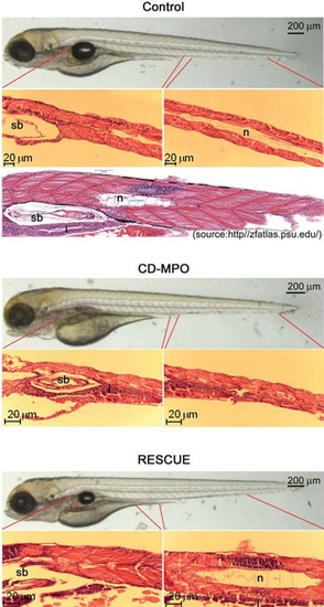
Histochemistry of the musculature in 4 dpf control, CD– MPO and rescue-CD zebrafish 4 dpf zebrafish larvae arising from zygotes injected with control or CD–MPO or CD–MPO plus rescue-CD mRNA were subjected to longitudinal sectioning and H&E staining of the musculature. Only zebrafish with an apparent ‘quasi-normal’ phenotype were analysed. A selection of images from three independent experiments is shown. The longitudinal section of wild-type larvae at the same age (http//zfatlas.psu.edu/) is reported for reference. Symbols: n, notochord; sb, swim bladder. |
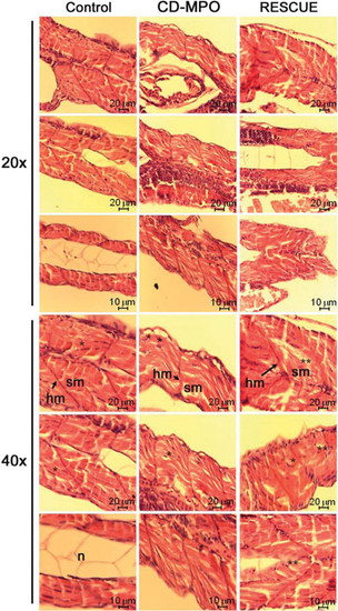
Particulars of the musculature of 4 dpf control, CD–MPO and rescue-CD zebrafish The most cranial, trunk and caudal portions of the skeletal musculature of the zebrafish described in Figure 4 were imaged at a high magnification as indicated. Symbols: hm, horizontal myoseptum; n, notochord; sm, somitic muscle.*syncytial nucleus; **eccentric nucleus. PHENOTYPE:
|

Unillustrated author statements PHENOTYPE:
|