
Image
|
Figure Caption
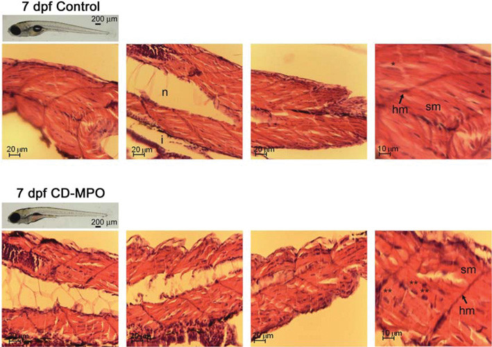
Fig. 2
Histochemistry of the musculature in 7 dpf control and CD–MPO zebrafish
Control and CD–MPO-injected zebrafish at 7 dpf showing an apparent ‘quasi-normal’ phenotype were subjected to longitudinal sectioning and H&E staining of the musculature. A selection of images from three independent experiments is shown. Symbols: hm, horizontal myoseptum; i, intestine; n, notochord; sm, somitic muscle.*syncytial nucleus; **eccentric nucleus.
Figure Data
Acknowledgments
This image is the copyrighted work of the attributed author or publisher, and
ZFIN has permission only to display this image to its users.
Additional permissions should be obtained from the applicable author or publisher of the image.
Full text @ Biosci. Rep.