
- Title
-
Evaluation of 14 organic solvents and carriers for screening applications in zebrafish embryos and larvae
- Authors
- Maes, J., Verlooy, L., Buenafe, O.E., de Witte, P.A., Esguerra, C.V., and Crawford, A.D.
- Source
- Full text @ PLoS One

Teratogenic effects of solvents within the first 24 hours post-fertilization. Typical results are shown. Embryos at 1 dpf treated for the previous 24 hours with a, embryo medium (control) or b, 1% DMF (from 4-cell stage), c, 1% DMF (from 4 hpf), d, 0.5% albumin (from 4-cell stage), e, 1.5% acetonitrile (from 4 hpf), and f, 2.5% glycerol (from 4-cell stage). b, embryos treated from 4-cell stage with 1% DMF display severe epiboly defects. The eyes (red arrow) and some somites are still visible (blue arrow), but most other structures are no longer clearly discernable. Massive cell death is also present surrounding the yolk (black asterisk). c, 1% DMF applied from 4 hpf onwards no longer inhibits epiboly. However, embryos display body curvature and shortening, loss of ventral tail fin (green arrow), no clear brain segmentation (black arrow), pericardial edema and a widened choroid fissure (orange arrow). d, embryos treated with 0.5% albumin are morphologically normal but display a significant delay in development (up to 9 hours) relative to control. e, 1.5% acetonitrile-treated embryos display anterior-posterior axis truncation, body curvature, microcephaly, lack of a midbrain-hindbrain boundary, incomplete pigmentation in the eyes, trunk and tail, ill-defined somitic boundaries, a bumpy notochord and ‘roughened’ epidermis and fin structures. f, embryos treated with 2.5% glycerol display anterior-posterior axis truncation with no extended tail, u-shaped somites, and cardia bifida (purple arrows). With the exception of embryo in panel b, which is oriented with anterior to the top, dorsal to the right, all other embryos are depicted with anterior to the left, dorsal to the top. |
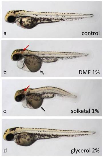
Teratogenic effects of solvents between 24 and 48 hours post-fertilization. Embryos at 2 dpf (a, b, c, d) treated for the previous 24 hours with a, embryo medium, b, 1% DMF, c, 1% solketal or d, 2% glycerol. Embryos in b, c, and d display no or very slow blood circulation as well as no touch response (all were immotile). Treatment with 1% DMF or 1% solketal also resulted in brain hemorrhaging (red arrows), pericardial edemas, yolk necrosis (black arrows) and shortening along the AP axis. d, Other than the observed blood flow defects, embryos treated with 2% glycerol appeared morphologically normal. All embryos are depicted with anterior to the left, dorsal to the top. |

Teratogenic effects of solvents between 72 and 96 hours post-fertilization. Zebrafish embryos at 4 dpf, treated for the previous 24 hours with a, embryo medium, or b and c, 2.5% DMSO. DMSO-treated larvae display either smaller or no swim bladders (red arrows) and necrosis in the yolk area surrounding the internal organs (white asterisk). All embryos are depicted with anterior to the left, dorsal to the top. |