
- Title
-
Mycobacterium marinum Causes a Latent Infection that Can Be Reactivated by Gamma Irradiation in Adult Zebrafish
- Authors
- Parikka, M., Hammarén, M.M., Harjula, S.K., Halfpenny, N.J., Oksanen, K.E., Lahtinen, M.J., Pajula, E.T., Iivanainen, A., Pesu, M., and Rämet, M.
- Source
- Full text @ PLoS Pathog.

ZFIN is incorporating published figure images and captions as part of an ongoing project. Figures from some publications have not yet been curated, or are not available for display because of copyright restrictions. PHENOTYPE:
|
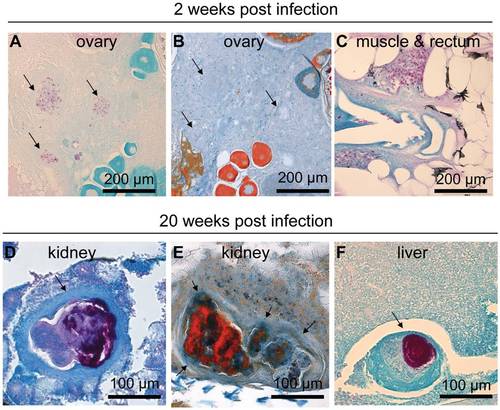
M. marinum induces the formation of granulomas that mature into well-defined structures during an infection. In fish infected with a low dose (34±15 cfu) of M. marinum, Ziehl-Neelsen staining at 2 wpi commonly reveals areas with free bacteria (C). Some slightly better formed and restricted areas containing bacteria, here referred to as early granulomas, are also seen (A), but as shown in (B) trichrome staining of the adjacent slide, encapsulation around the mycobacterial lesions is absent at the early stage of infection. At 20 weeks, fish that have survived have mature granulomas (D–F) many of which are multicentric surrounded by a fibrous capsule (D&E). (E) Trichrome staining shows the fibrous capsule in blue (F). The amount of bacteria inside granulomas has increased from the earliest time-points. PHENOTYPE:
|
|
ZFIN is incorporating published figure images and captions as part of an ongoing project. Figures from some publications have not yet been curated, or are not available for display because of copyright restrictions. |
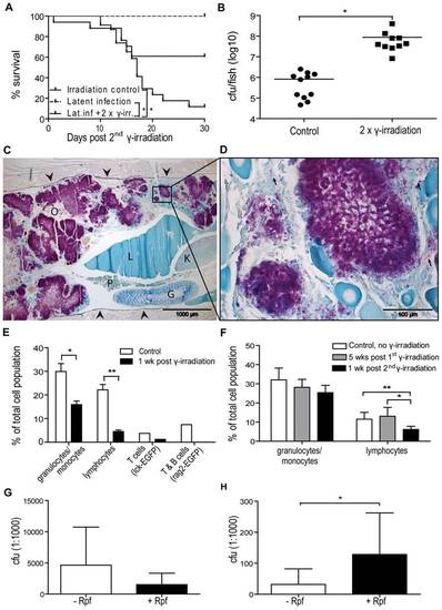
Gamma irradiation induces reactivation resulting in increased mortality due to uncontrolled growth of mycobacteria. (A–C) Zebrafish (n = 17) with a latent M. marinum infection were irradiated twice with 25 Gy with one month between the irradiations. Twice irradiated, non-infected zebrafish (n = 23) as well as zebrafish with a latent infection (n = 14) were included as controls. (A) Survival was followed for 30 days after the second dose. *P<0.05. (B) During this period, moribund or recently dead fish were collected 15–22 days after the second radiation dose. Bacterial loads were compared with those of similarly infected, non-irradiated control fish that were collected at the end-point of the experiment. *P<0.05 (C&D) A representative Ziehl-Neelsen stained sample from a reactivated fish showing large numbers of free mycobacteria (purple areas) in the zebrafish body cavity (C). The sides of the body cavity are marked with arrowheads O = ovary, P = pancreas, L = liver, G = gut, K = kidney. (D) A picture taken with a higher magnification showing individual rods (few examples pointed out with arrows). (E) Four groups of 4 adult zebrafish (1 rag2-gfp, 1 lck-gfp and 2 wild-type groups) were γ-irradiated with 25 Gy. Similar control groups were left untreated. Kidneys were collected 8 d post irradiation, pooled and analyzed by FCM. FSC-SSC -plots were gated based on cell size and granularity as described in [56] (gates shown in Figure S3) to assess the effect of irradiation on leukocyte populations. *P<0.05. For further verification of the effect of radiation on lymphocytes, a GFP gate was used for the rag2 and lck groups expressing GFP in B and T cells, or T cells, respectively. (F) Adult non-infected wt zebrafish were irradiated with 25 Gy once (grey bars) (n = 3) or twice (n = 7) (black bars) with one month between the doses. Leukocyte recovery and re-depletion were assessed by FCM. Non-irradiated fish (n = 4) were used as controls. *P<0.05 (G) Fish with a latent infection (n = 7) were irradiated twice with 25 Gy with one month between the doses and plated +/- Rpf for 18 d after the second radiation dose. (H) Fish (n = 6) with a latent infection were plated +/- Rpf. PHENOTYPE:
|