
- Title
-
STARS Is Essential to Maintain Cardiac Development and Function In Vivo via a SRF Pathway
- Authors
- Chong, N.W., Koekemoer, A.L., Ounzain, S., Samani, N.J., Shin, J.T., and Shaw, S.Y.
- Source
- Full text @ PLoS One
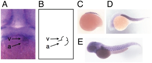
Whole mount in situ expression of zSTARS in zebrafish embryos. A. Frontal view of 48 hpf embryo showing zSTARS expression in ventricle (v) and atrium (a). B. Schematic depicting position of ventricular (v) and atrium (a) in the view from part A. C. Expression in somites at 16 somite stage (17 hpf). D. and E. Expression in somites and central nervous system at 24 hpf (D) and 48 hpf (E). EXPRESSION / LABELING:
|
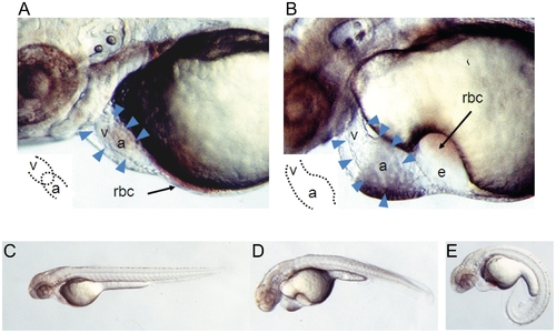
Developmental phenotype of morpholino-induced knockdown of zSTARS: lateral views of 56 hpf embryos. A. Embryo injected with control morpholino (with 5 mismatches). The cardiac silhouette is demarcated by arrowheads. The heart tube is looped so that the ventricle (v) and atrium (a) are closely apposed (inset). Circulating red blood cells (rbc) are visible in a thin rim along the inferior aspect of the yolk and within the heart. B. Embryo injected with zSTARS morpholino. The heart tube is unlooped so that the ventricle (v) and atrium (a) are co-linear, with atrial dilation (inset). There is significant edema in the pericardium and over the yolk, with stasis of red blood cells (rbc) over the yolk. C. Lateral view of entire 56 hpf embryo following injection of control, mismatch morpholino. D. and E. 56 hpf embryos showing representative phenotypes of zSTARS morpholino injection. PHENOTYPE:
|
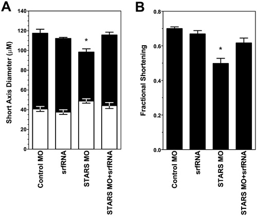
Ventricular function and dimensions based on quantitative analysis of high-speed video microscopy. A. Ventricular dimensions at end-diastole (black bars) and end-systole (white bars). Conditions are identical to those in part a. Values plotted are mean (n = 4 embryos) ± standard deviation. Asterisk (*) denotes statistically significant difference by ANOVA. B. Ventricular fractional shortening observed after injection of: control mismatched morpholino (MM MO), zSTARS morpholino + srf mRNA (MO + SRF), srf mRNA only (SRF only), or zSTARS morpholino only (MO only). Values plotted are mean (n = 4 embryos) ± standard deviation. Asterisk (*) denotes statistically significant difference by ANOVA. PHENOTYPE:
|
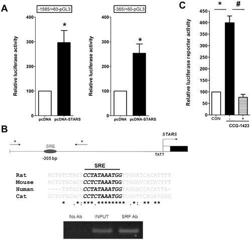
Binding of SRF to the STARS promoter. A. STARS expression activates reporter gene constructs containing the conserved serum response element (SRE). Luciferase activity is shown for two constructs upstream of the STARS transcription start site. B. Chromatin immunoprecipitation assays were performed with formaldehyde cross-linked chromatin isolated from feline adult cardiomyocytes. Asterisk (*) denotes PCR primer locations. Immunoprecipitations were performed without primary antibody (No Ab) as a negative control, with anti-SRF antibody. Input DNA is also shown as a positive control. Similar results were observed in four independent experiments. C. The SRF inhibitor CCG-1423 (1 µM) abolished STARS −365/+60 promoter-reporter activity in H9c2 cells (n = 3 experiments, in triplicates). |
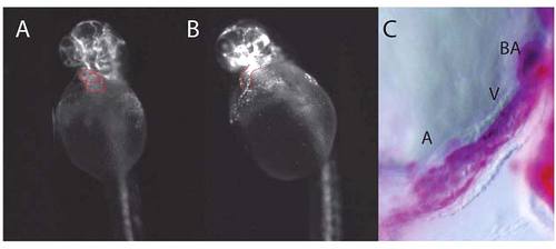
Tg(FLK:G-RFP) embryos were injected with MO at the 1 cell stage and allowed to develop under standard conditions. [2], and fluorescent and bright-field images of 48 hpf zebrafish are shown. Panel A: Tg(FLK:G-RFP) injected with mismatch MO showing normal cardiac looping. Panel B: Tg(FLK:G-RFP) injected with STARS MO showing defective looping. Panel C: Bright-field image of the heart following digital subtraction of fluorescent Tg(FLK:G-RFP) image: this highlights the cardiac lumen through the atrium (A), ventricle (V) and the bulbus arteriosus (BA), showing that the outflow tract is unobstructed. |
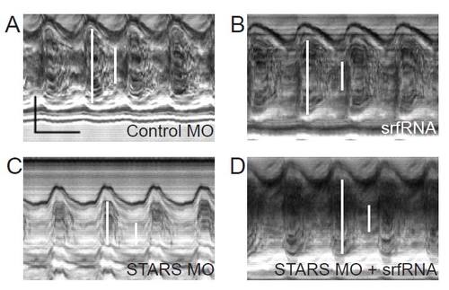
M-mode images of zebrafish ventricles. Fertilized zebrafish oocytes were injected with (A) control morpholino, (B) in vitro transcribed mRNA for srf(srfRNA), (C) morpholino targeting STARS mRNA (moSTARS) or (D) moSTARS and srfRNA and allowed to develop under standard conditions. High speed video images of ventricles were obtained at 48 hpf and motion mode (m-mode) images obtained at 125 frames per second as described [3]. In these frames, the vertical dimension displays the image across a single scan line over time in the horizontal dimension. Ventricular short axis diameter (white bars) and myocardial thickness were measured at end diastole and end systole in each condition. In panel (A), the vertical black bar denotes 50 microns and the horizontal bar denotes 500 msec. PHENOTYPE:
|

Unillustrated author statements EXPRESSION / LABELING:
|