
|
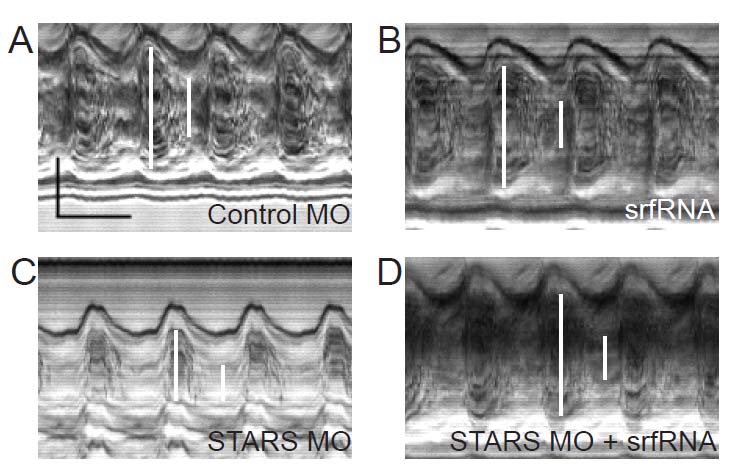
Fig. S3
M-mode images of zebrafish ventricles.
Fertilized zebrafish oocytes were injected with (A) control morpholino, (B) in vitro transcribed mRNA for srf(srfRNA), (C) morpholino targeting STARS mRNA (moSTARS) or (D) moSTARS and srfRNA and allowed to develop under standard conditions. High speed video images of ventricles were obtained at 48 hpf and motion mode (m-mode) images obtained at 125 frames per second as described [3]. In these frames, the vertical dimension displays the image across a single scan line over time in the horizontal dimension. Ventricular short axis diameter (white bars) and myocardial thickness were measured at end diastole and end systole in each condition. In panel (A), the vertical black bar denotes 50 microns and the horizontal bar denotes 500 msec.