
- Title
-
Persistent oxytetracycline exposure induces an inflammatory process that improves regenerative capacity in zebrafish larvae
- Authors
- Barros-Becker, F., Romero, J., Pulgar, A., and Feijoo, C.G.
- Source
- Full text @ PLoS One
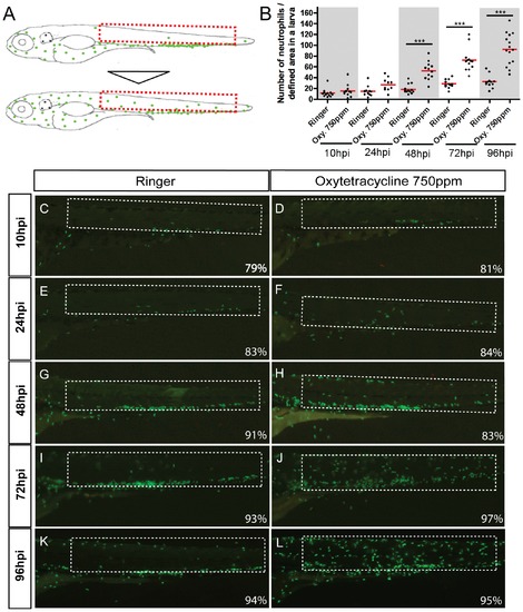
Exposure to oxytetracycline triggered an inflammation process. Incubation of Tg(mpx:GFP) transgenic larvae (that express GFP exclusively in neutrophils) in oxytetracycline 750 ppm induce a progressive migration of neutrophils from to the caudal hematopoietic tissue (CHT) to the entire tail. (A) Scheme of neutrophils localization in control and experimental larvae. (B) Quantification of neutrophils migration into the selected area. (C–L) Close up of the tail of control and experimental embryos at 10 hour post incubation (hpi), 24hpi, 48hpi, 72hpi and 96hpi. At 10hpi (C, D) and 24hpi (E, F) we did not detect any significant neutrophils migration. Later, at 48hpi (G, H) a discrete number of neutrophils migrate from de CHT to the tail. At 72hpi (I, J) and 96hpi (K, L), the presence of the GFP positive cells in the tail becomes clearly evident. The numbers of larvae that presented the phenotype shown is expressed as a percentage. For all experiments, at least 13 larvae were used for each condition. *** p<0.001. |
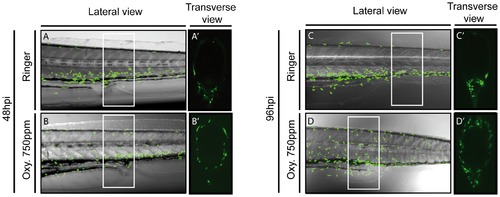
Oxytetracycline exposure induces neutrophils migration to superficial tissues. We analyzed the Tg(mpx:GFP) transgenic line, by confocal microscopy, detecting that in oxytetracycline exposure larvae neutrophils migrated to superficial tissue. (A–D) lateral view, (A2–D2) transverse view. Transverse views show a section of a larval tail of approximately 250 μm (white rectangle in lateral view). |
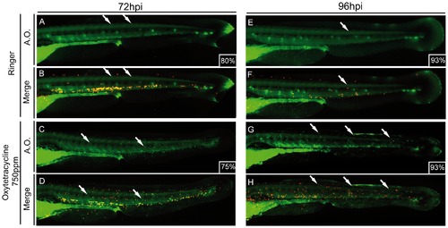
Cell death is detected in advanced steps of oxytetracycline induced inflammation. 48hpf Tg(LyzC:DsRED2) transgenic larvae were incubated for 72 hrs (A–D) or 96 hrs (E–H) in oxytetracycline 750 ppm followed by an acridine orange stain to address cell death. Experimental larvae showed higher level of cell death (white arrows) compared to control at both time analyzed. The numbers of larvae that presented the phenotype shown is expressed as a percentage. |
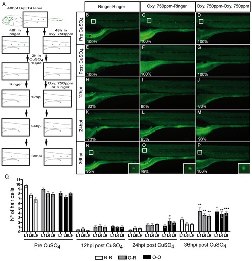
Oxytetracycline induced inflammation increase regeneration capacity. (A) Scheme of the regeneration assay developed. Incubation of SqET4 transgenic larvae (that express GFP in neuromast hair cells, HC) in oxytetracycline 750 ppm increases the rate of GFP/hair cells appearance. (B–D) HC before copper treatment. (E–G) CuSO4 treatment eliminates all GFP positive cells. (H–M, Q) Although we observe a tendency, there is no significant difference in the number of regenerated HC. (N–Q) At 36hpi the regeneration capacity of HC is significantly increased on oxytetracycline exposed larvae. (Q) The quantification of hair cells was performed in three neuromast; L1, L6 and L9. White box indicate L1 neuromast hair cells. The numbers of larvae that presented the phenotype shown is expressed as a percentage. * p<0.05, ** p<0.01, ***p<0.001. |
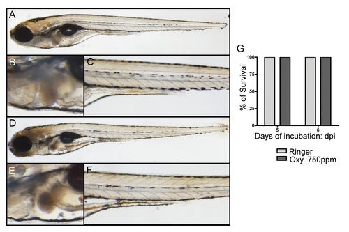
6 days of oxytetracycline exposure does not produce mortality nor adverse effects on larvae. Larvae were incubated during 120 hrs and 144 hrs to ensure that the treatment with oxytetracycline does not produce any detrimental effects on larvae. We did not found phenotypic effects such cerebral edema, bending of the tail (A, D), pericardial edema, abnormal heart function (B, E), or any somite malformation (C, F). No change in survival rate was detected at the time analyzed (G). |