
- Title
-
Identification and Analysis of Conserved cis-Regulatory Regions of the MEIS1 Gene
- Authors
- Royo, J.L., Bessa, J., Hidalgo, C., Fernández-Miñán, A., Tena, J.J., Roncero, Y., Gómez-Skarmeta, J.L., and Casares, F.
- Source
- Full text @ PLoS One
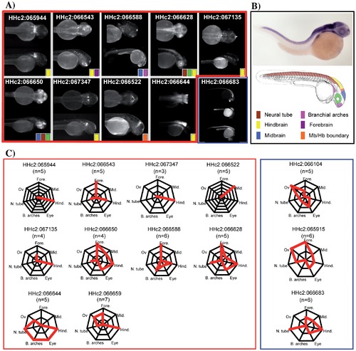
Enhancer activities from the HCNRs recapitulate endogenous meis1 expression pattern. A) Dorsal and lateral views of a representative founder illustrate the expression pattern of each HCNR. B) Picture of a meis1 in situ hybridization of a 30 hpf embryo illustrates the different expression territories. A color-based schema of a zebrafish embryo highlights the different domains where GFP expression was found during the study. C) Diagrams of patterns of expressions of highly penetrant enhancers (boxed in red) and of non-tissue specific enhancers (boxed in blue). In each diagram, the number of founder lines showing expression in each particular body structure is represented by the red lines. For example, of three lines of HHc2:066543, all three showed forebrain expression, and additionally, two of them drove expression in the hindbrain while the remaining one in the neural tube. In situ hybridization picture is from ZFIN [16]. EXPRESSION / LABELING:
|
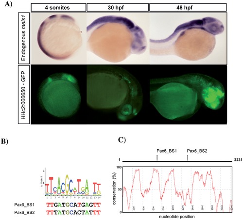
HCNR sequence analysis suggests novel meis1 upstream regulators. (A) meis1 transcription, detected by in situ hybridization (upper panel) compared to GFP expression, driven by the HHc2:066650-GFP transgenic line (lower panel) at 12 (left), 24 (middle) and 30 hpf (right). Side views are shown. HHc2:066650 drives GFP expression in the eye primordium, the early hindbrain, the retina, the optic tectum and the olfactory bulb, which are meis1 expression domains. B) The analysis of the potential binding sites predicted by Jaspar highlights the presence of three potential Pax6 sites. Two of these sites contain a high degree of similarity with the consensus. (C) Pax6_Binding_Site1 also mapped to a microisland of ultraconservation when human and zebrafish orthologous regions were aligned. EXPRESSION / LABELING:
PHENOTYPE:
|
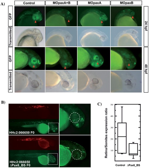
Pax6 regulates the enhancer activity of HHc2:066650. A) Representative pictures of transgenic zebrafish embryos at 24 and 48 hpf, corresponding to HHc2:066650 F2 offspring injected with MOpaxA, MOpaxB, or a mixture of both. The red arrow points to the retina, where a strong decrease in the GFP levels is observed after morpholino treatment. A control mopholino-injected individual is shown for comparison. B) Representative mosaic embryos injected with HHc2:066650 construct with/without the two candidate Pax6 binding sites. Both the GFP and RFP channels are included. The boxed region is magnified on the right. The dashed circle delineates the eye. (C) GFP expression was measured in the eye (area marked by the dashed circle in (B)) of wild type and Δpax6_BS version of HHc2:066650 mosaic embryos (n = 9 and 11 embryos, respectively), and normalized with their respective muscle RFP expression. Enhancer signal significantly decreased in the mutant version of HHc2:066650 (p = 0.018, Mann Whitney test). The box plots contains a central rectangle, which spans from the first quartile to the third quartile. A segment inside the rectangle shows the median and whiskers above and below the box show the locations of the minimum and maximum. |
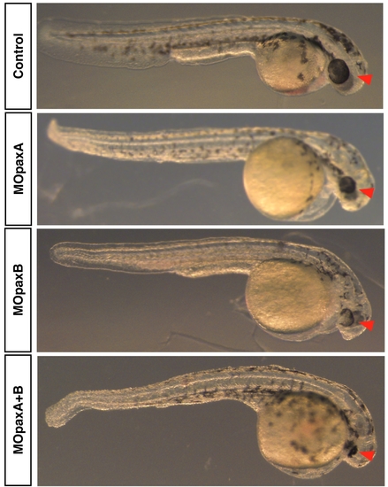
Morpholinos against Pax6a and Pax6b affect eye development. Representative pictures of 48 hpf wild type zebrafish embryos after different MO injection. The microphthalmia observed among morphants (red arrow) confirmed the functionality of the morpholinos. No effects on control morpholino injected animals were detected. |