
- Title
-
The Cx43-like Connexin Protein Cx40.8 Is Differentially Localized during Fin Ontogeny and Fin Regeneration
- Authors
- Gerhart, S.V., Eble, D.M., Burger, R.M., Oline, S.N., Vacaru, A., Sadler, K.C., Jefferis, R., and Iovine, M.K.
- Source
- Full text @ PLoS One
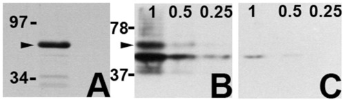
The Cx40.8 antibody is specific. (A) An antibody raised against a Cx40.8 carboxy-terminal peptide detects a single major band from regenerating fin lysates (arrowhead). Non-competed (B) and competed (C) anti-Cx40.8 antibody was used to detect bacterially expressed GST-Cx40.8CT. Decreasing volumes of a concentrated lysate were loaded in each lane on two identical gels. When the antibody was pre-incubated with the Cx40.8 target sequence (i.e. competed), antibody binding was reduced in all lanes compared with non-competed antibody. Arrowhead points to the full length product. Bands at molecular weights smaller than the full-length fusion protein represent degradation products. |
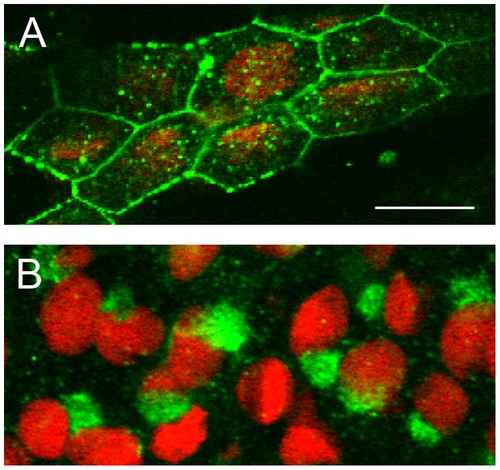
Cx40.8 is localized to different subcellular compartments during ontogeny and regeneration. (A) During ontogeny, Cx40.8 immunofluorescence (green) on whole fins counterstained with propidium iodide (red) shows that Cx40.8 locates to the plasma membrane and vesicles. (B) During regeneration, Cx40.8 is intracellular and staining is consistent with the Golgi apparatus. |
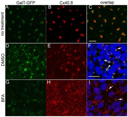
Cx40.8 co-localizes with the GalT-GFP transgene found in the Golgi during regeneration. (A–C) Co-localization in untreated regenerating fins. (D–F). Co-localization in regenerating fins treated with 0.1% DMSO carrier. (G–I) Fins injected with10 µg/ml BFA/0.1% BFA to disrupt the Golgi show dispersal of both Cx40.8 and GalT-GFP signals. In D-I, nuclei (blue) are stained with TO-PRO3 detected in the far red channel. Arrows point to areas of overlap in F (intact Golgi) and I (remnants of intact Golgi). Scale bars in C and F, 10 μm. |
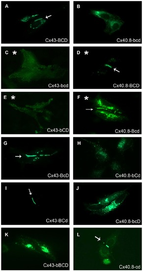
The Cx40.8-b domain is responsible for the intracellular localization of Cx40.8. HeLa cells were singly transfected with EGFP fusions of each construct. (A) Cx43-BCD-EGFP, (B) Cx40.8-bcd-EGFP, (C) Cx43-bcd-EGFP, (D) Cx40.8-BCD-EGFP, (E) Cx43-bCD-EGFP, (F) Cx40.8-Bcd-EGFP, (G) Cx43-BcD-EGFP, (H) Cx40.8-bCd-EGFP, (I) Cx43-BCd-EGFP, (J) Cx40.8-bcD-EGFP, (K) Cx43-bBCD-EGFP, (L) Cx40.8-cd-EGFP. Asterisks denote constructs in which the localization depends on a component of the carboxy terminus. Arrows identify gap junction plaques. |
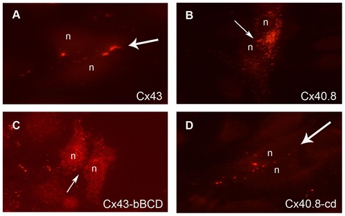
Untagged Cx43 and Cx40.8 constructs behave similarly as GFP tagged constructs in transiently transfected HeLa cells. (A) Cx43 localizes to the plasma membrane. (B) Cx40.8 is retained intracellularly. (C) Cx43-bBCD is retained intracellularly. (D) Cx40.8-cd localizes to the plasma membrane. The Cx43 antibody [12] was used to detect untagged Cx43 and untagged Cx43-bBCD. The Cx40.8 antibody was used to detect untagged Cx40.8 and untagged Cx40.8-cd. Flared arrows identify gap junction plaques located at the plasma membrane; Plain arrows identify the plasma membrane in the absence of gap junction plaques; n, nucleus. |
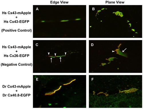
Cx43-mApple and Cx40.8-EGFP co-assemble in common gap junction channels. High resolution fluorescence microscopy was used to provide evidence for co-association of Cx43-mApple and Cx40.8-EGFP in common gap junction channels. Constructs that were co-transfected in HeLa cells are indicated to the left of the panels (A, B) Homo sapiens (Hs) Cx43-mApple + Hs Cx43-EGFP show uniformly yellow plaques, suggesting co-asociation. (C, D) Hs Cx43-mApple + Hs Cx26-EGFP show discrete green and red domains, revealing a lack of co-association. Arrows indicate green Hs Cx26-EGFP localization and arrowheads indicate red Hs Cx43-mApple localization. (E, F) Danio rerio (Dr) Cx43-mApple + Dr Cx40.8-EGFP show uniform yellow distribution. |
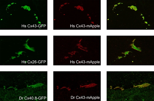
Cx43-mApple and Cx40.8-EGFP co-assemble in common gap junction channels. High resolution fluorescence microscopy was used to provide evidence for co-association of Cx43-mApple and Cx40.8-EGFP in common gap junction channels. Plane views are shown. Top: In HeLa cells co-transfected with Hs-Cx43-EGFP and Hs-Cx43-mApple, both green and red plaques completely overlap. Middle: In HeLa cells co-transfected with Hs-Cx26-GFP and Hs-Cx43-mApple, discrete domains of green and red plaques are observed. Bottom: In HeLa cells co-transfected with Dr-Cx40.8-EGFP and Dr-Cx43-mApple, both green and red plaques completely overlap. |