
- Title
-
Targeted knock-down of a structurally atypical zebrafish 12S-lipoxygenase leads to severe impairment of embryonic development
- Authors
- Haas, U., Raschperger, E., Hamberg, M., Samuelsson, B., Tryggvason, K., and Haeggström, J.Z.
- Source
- Full text @ Proc. Natl. Acad. Sci. USA
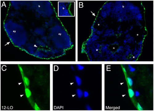
Expression pattern of zf12-LO protein in zf embryos: Immunohistochemical staining of zf12-LO was performed on transversal sections from a two dpf embryo. Nuclear expression of zf12-LO was detected in the skin epithelium that surrounds the embryo (A and B, arrows) as well as the yolk sac. zf12-LO was also detected in the epithelial lining of the stomodeum (A, arrowhead) and the pharyngeal pouches (B, asterisks). Unspecific staining of the zf12-LO antibody is shown in insert in A and in the notochord (B). The notochord (n), brain (b), eyes (ey), and ear (e) are indicated for orientation. Epithelial cells at higher magnification (C-E) show nuclear localization of zf12-LO as assessed by colocalization (arrow heads) of zf12-LO immunostaining and DAPI staining (blue). EXPRESSION / LABELING:
|
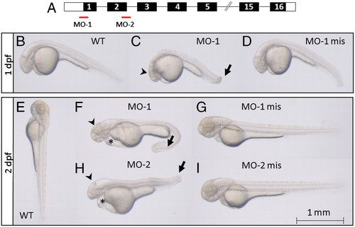
Knock-down of zf12-LO leads to abnormal development of the head and tail as well as yolk sac and pericardial edema. (A) Two MOs, one complementary to the region of the ATG site blocking translation (MO-1) and another corresponding to the end of exon-2/ start of intron-2/3 blocking the correct splicing of exon-2 (MO-2) were designed. The exons of the zf12-LO gene, encoding the zf12-LO protein, are shown in black boxes and the 5′UTR and 3′UTR regions are marked with white boxes (not drawn to scale). Embryos were visualized by regular light microscopy at one dpf (B-D) and two dpf (E-I). Wild type (B) as well as MO-1 mis treated embryos (D) developed normally whereas injection of MO-1 (C) resulted in retarded development of the head (arrowhead) and a bent tail (arrow) already after one dpf. The phenotype was more pronounced in MO-1 morphants at two dpf (F). The head was less developed (arrowhead), the tail was strongly bent (arrow) and pericardial edema as well as edema around the yolk sac appeared (asterisk). A similar phenotype was observed in MO-2 treated embryos at two dpf (H). In contrast, WT (E) as well as embryos treated with MO-1 mis and MO-2 mis (G and I) developed normally. Animals shown are representative embryos for the indicated time points. |
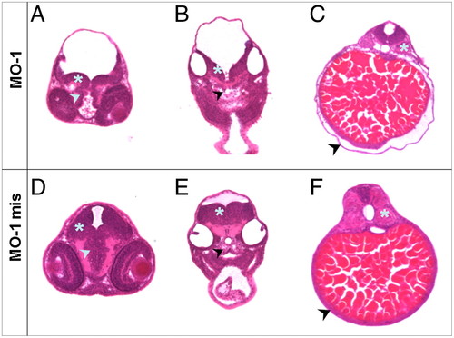
Transversal sections through different layers of embryos at two dpf treated with MO-1 and MO-1 mis. Embryos were embedded in plastic, 10 µm transversal sections were prepared and counterstained with Hematoxylin/Eosin. The regions of eye/brain (A and D), ear/heart (B and E) and kidney/gut (C and F) were analyzed. (A) MO-1 treated embryos display a delayed development of the Mesencephalon (asterisk) as well as Diencephalon (arrowhead) compared to MO-1 mis treated embryos (D). (B) The decelerated development of the brain was also detectable in the Myelencephalon region (asterisk) in MO-1 morphants combined with an unstructured composition of the basal plate (arrowhead). (E) MO-1 mis treated embryos showed a normally developed Myelencephalon and a well constructed basal plate. (C) In the area of the kidney/gut, an edema located around the yolk sac was observed in MO-1 morphants (arrowhead). Furthermore, an unstructured composition of the somites was visible (asterisk). (F) These alterations were not observed in MO-1 mis treated embryos. Sections shown are from representative embryos for the indicated time points. PHENOTYPE:
|

Zf12-LO is required for normal eye development. Sections of zf embryos were prepared and stained with Hematoxylin/Eosin. Morphological comparison of zf12-LO morphants (treated with MO-2) and controls (treated with MO-2 mis) was carried out with light microscopy at two dpf. Injection of MO-2 results in developmental eye abnormalities including loss of retinal layered structures and lens crystalline. RPE; retinal pigmented epithelium, RCL; rods and cones layer, OPL; outer plexiform layer, BCL; bipolar cell layer, ACL; amacrine cell layer, IPL; inner plexiform layer, GCL; ganglion cell layer. Similar results were obtained with zf12-LO morphants treated with MO-1. PHENOTYPE:
|

Unillustrated author statements PHENOTYPE:
|