
|
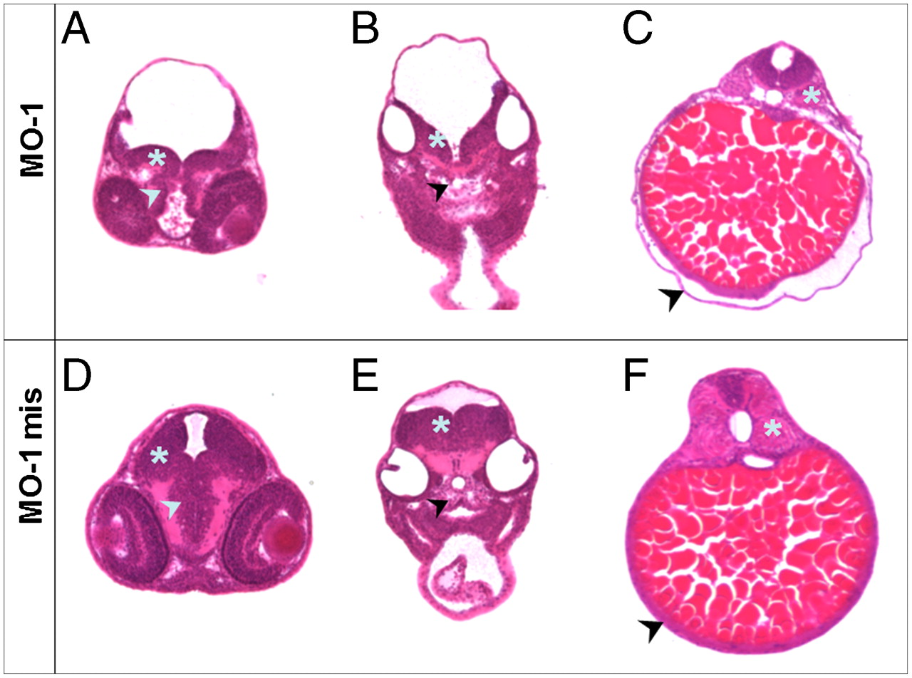
Fig. 4
Transversal sections through different layers of embryos at two dpf treated with MO-1 and MO-1 mis. Embryos were embedded in plastic, 10 µm transversal sections were prepared and counterstained with Hematoxylin/Eosin. The regions of eye/brain (A and D), ear/heart (B and E) and kidney/gut (C and F) were analyzed. (A) MO-1 treated embryos display a delayed development of the Mesencephalon (asterisk) as well as Diencephalon (arrowhead) compared to MO-1 mis treated embryos (D). (B) The decelerated development of the brain was also detectable in the Myelencephalon region (asterisk) in MO-1 morphants combined with an unstructured composition of the basal plate (arrowhead). (E) MO-1 mis treated embryos showed a normally developed Myelencephalon and a well constructed basal plate. (C) In the area of the kidney/gut, an edema located around the yolk sac was observed in MO-1 morphants (arrowhead). Furthermore, an unstructured composition of the somites was visible (asterisk). (F) These alterations were not observed in MO-1 mis treated embryos. Sections shown are from representative embryos for the indicated time points.