
- Title
-
Ancestral network module regulating prdm1 expression in the lamprey neural plate border
- Authors
- Nikitina, N., Tong, L., and Bronner, M.E.
- Source
- Full text @ Dev. Dyn.

Expression pattern of prdm1 in sea lamprey embryos. A–N: Whole-mount in situ hybridization with the full-length lamprey prdm1 probe. Orientation: anterior is at the top in dorsal views, or in the top right-hand corner in side views. L2,O: A 12-micron sections through the stained embryos. A: Dorsal view of an embryonic day (E) 4.0 embryo, showing prdm1 transcripts expressed uniformly throughout the ectoderm and the neural plate border, but absent from the neural plate. B: Side view of the same embryo. C: Dorsal view of the embryo in (A) with its ectoderm removed, demonstrating that prdm1 is also expressed in the paraxial mesoderm at this stage. D,E: Starting at E4.5 (D), high levels of prdm1 are expressed in the neural plate border (white arrows) and in the preplacodal domain (black arrowhead), and lower levels in the ectoderm. E: A similar pattern of prdm1 expression is seen 6 hours later at E4.75. F,G: Dorsal (F) and side (G) view of the same E5.0 embryo, showing strong expression in the premigratory neural crest (white arrow). H: Embryo at E5.5. prdm1 is turned off in the neural crest as it starts to migrate. I: Lateral view of an E6.5 embryo. prdm1 transcripts are no longer seen in the anterior dorsal neural tube. J,K: Dorsal (J) and side (K) view of an E7.5 embryo. prdm1 is expressed in the trunk dorsal neural tube, in the branchial basket and in somites. L: E8.5 embryo showing prdm1 expression in the branchial arches. M: Side view of the head of an E10.5 embryo, showing prdm1 expression in the lips and lens (lp) and otic placodes (op). N: Side view of E20 embryo, showing prdm1 expression in the cartilage of the branchial arches. (L2) Transverse section through the head of the E8.5 embryo in (L) showing prdm1 expression in the endoderm and the lateral region of the neural crest (black arrowhead, compare with panel O). O: Section through an E10.5 embryo stained for Col2a2, which at that stage is expressed in the neural crest components of the branchial arches. Black arrow marks the lateral component of the neural crest that corresponds to the prdm1 expressing cells in (L2). m, paraxial mesoderm; pnc, premigratory neural crest; op, otic placode; lp, lens placode. White arrows indicate neural plate border, black arrowheads, preplacodal domain. |
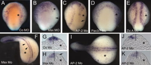
Expression of prdm1 in the neural plate border is regulated by AP-2 and Msx A. prdm1 is detected by in situ hybridization in morpholino-injected embryos. A–E: Top row: Embryonic day (E) 4.5 embryos injected with control (A) Msx A (B), AP-2 (C), Pax3/7 (D), and Zic A (E) morpholinos. All embryos are shown in dorsal view with anterior facing up. In all embryos, morpholino was incorporated into the right side (marked with an asterisk). Arrows in (B) and (C) indicate loss of prdm1 expression on the injected side. F: MsxA-morpholino-injected embryo at E8.5, showing loss of prdm1 expression in somites and reduction of expression in half of the neural tube (arrowheads). H: Section through the embryo in (F) showing loss of prdm1 expression in one half of the neural tube (arrow). G: Section through an E8.5 embryo injected with the control morpholino. I: E7.5 embryo injected with AP-2 morpholino, showing that prdm1 expression in the somites in not affected (asterisk marks the injected side). J,K: Sections through the embryo in panel I, showing loss of prdm1 expression on the injected side (asterisk). |
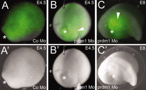
prdm1 Mo never incorporates in ectoderm. A–C: Fluorescent images showing incorporation of fluorescein isothiocyanate (FITC) -labeled morpholino (green). A2–C2: Brightfield images of the same embryos. A,A2: Embryonic day (E) 4.5 embryo injected with a control morpholino, showing incorporation of the morpholino throughout the dorsal ectoderm. The embryo is shown in dorsal view, with the anterior in the top right-hand corner. Asterisk denotes the position of the blastopore. B,B2: E4.5 embryo injected with prdm1 morpholino. Morpholino is incorporated in the mesoderm and endoderm (white arrowhead). The embryo is shown with the dorsal side facing up, and the posterior (blastopore, marked with an asterisk) facing toward the viewer. C,C2:prdm1 morpholino-injected embryo at E8. Orientation: the head is facing right, and the dorsal side is facing up. Asterisk denotes the abnormal position of the blastopore. |