
- Title
-
Hypoxia Impairs Primordial Germ Cell Migration in Zebrafish (Danio rerio) Embryos
- Authors
- Lo, K.H., Hui, M.N., Yu, R.M., Wu, R.S., and Cheng, S.H.
- Source
- Full text @ PLoS One
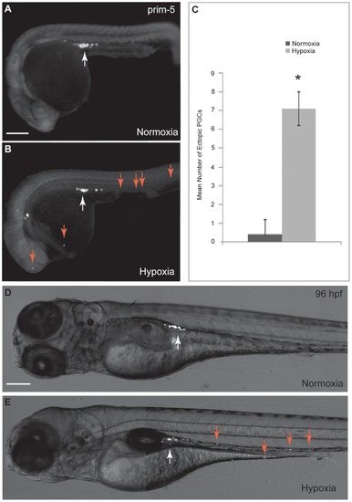
Hypoxia affects PGC migration in zebrafish embryos. Embryos were injected with GFP-nosl 32UTR mRNA as PGC marker. (A) PGCs in normoxic embryos migrated properly towards the genital ridge at prim-5 stage (white arrow). (B) PGC migration is affected by hypoxia as illustrated by presence of mis-migrated ectopic PGCs at prim-5 stage (red arrows). (C) Significantly greater number of mis-migrated ectopic PGCs was observed in hypoxic embryos as compared with normoxic embryos at prim-5 stage. (D) PGCs in normoxic embryos located at the genital ridge at 96 hpf. (E) Mis-migrated ectopic PGCs in yolk sac and in cranial region remained in the same position and failed to move towards the genital ridge at 96 hpf (red arrows). Scale bar: 200 μm. * denotes p<0.05. PHENOTYPE:
|
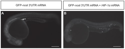
Effect of HIF-1α mRNA over-expression on PGC migration in zebrafish embryos. (A) Embryos injected with GFP-nosl 32UTR mRNA. (B) Embryos co-injected with GFP-nosl 32UTR mRNA and HIF-1α mRNA. HIF-1α over-expressed embryos showed mis-migrated ectopic PGCs in the head, yolk sac extension and caudal region. HIF-1α over-expressed embryos displayed similar phenotypes as embryos exposed to hypoxia. Scale bar: 200 μm. |
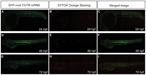
Hypoxia does not promote cell death in mis-migrated ectopic PGCs. (A–C) Embryos injected with GFP-nosl 32UTR mRNA were exposed to hypoxia and stained with SYTOX orange for dead cells. Mis-migrated ectopic PGCs (green) were not removed by apoptosis. The merged images show no co-localization between mis-migrated ectopic PGCs (green) and dead cells stained by STYOX Orange (red). |
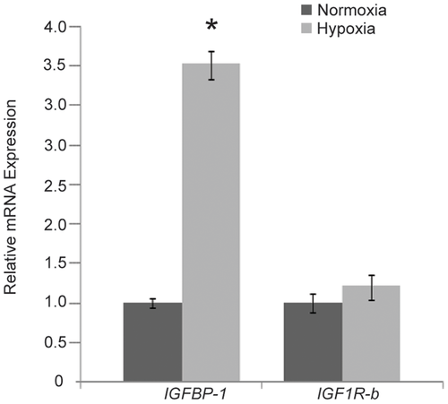
Hypoxia induces IGFBP-1 mRNA expression in zebrafish embryos at prim-5 stage. Expression levels of IGFBP-1 mRNA and IGF1R-b mRNA were quantified using real-time PCR and normalized against 18S rRNA. Data are the mean relative fold changes ± SE (n = 10) with respect to the control (normoxia) level (arbitrarily set to 1). Significantly higher IGFBP-1 mRNA expression was found in hypoxic embryos as compared with normoxic embryos. No significant difference in IGF1R-b mRNA expression was found between normoxic and hypoxic embryos. * denotes p<0.05. EXPRESSION / LABELING:
|
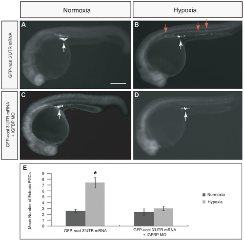
Effect of IGFBP-1 knockdown on PGC migration in zebrafish embryos. (A and B) Embryos were injected with GFP-nosl 32UTR mRNA alone and subsequently exposed to either normoxia or hypoxia. PGC migration was impaired in hypoxic embryos (B) as compared with normoxic embryos (A). (C and D) Embryos were co-injected with GFP-nosl 32UTR mRNA and IGFBP-1 MO and subsequently exposed to either normoxia or hypoxia. (D) PGC migration defect in hypoxic embryos was minimized by IGFBP-1 MO knockdown. Scale bar: 200 μm. (E) Mean number of mis-migrated ectopic PGCs in normoxia and hypoxia embryos injected with GFP-nosl 32UTR mRNA alone or co-injected with GFP-nosl 32UTR mRNA and IGFBP-1 MO. The mis-migrated ectopic PGCs phenotype could be rescued by IGFBP-1 MO knockdown as indicated by reduction in number of mis-migrated ectopic PGCs in hypoxic embryos. * denotes p<0.05. |