
- Title
-
Alkali-like myosin light chain-1 (myl1) is an early marker for differentiating fast muscle cells in zebrafish
- Authors
- Burguière, A.C., Nord, H., and von Hofsten, J.
- Source
- Full text @ Dev. Dyn.
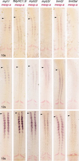
Expression pattern of early markers for fast-twitch muscles. Whole-mount in situ hybridization showing expression of myl1, fMyHC1.3, mylz2, mylz3, tnni2, and tnnt3a in embryos at 10-, 12-, and 15-somite stages. Expression of myl1, fMyHC1.3, mylz2, and tnni2 is detected in the paraxial mesoderm at 10-somite stage, mylz3 expression is first detected at 12-somite stage, and tnnt3a starts to be expressed at 15-somite stage. mesp-a (in red) labels somite primordia. Arrowheads indicate the earliest expressing somite. |

Expression of myl1 during early- to mid-somitogenesis. Expression of myl1 was detected in the paraxial mesoderm by whole mount in situ hybridization from the 8-somite stage. A: myl1 (blue) and smyhc1 (red) expression in flat-mounted and (A2) transverse section in an 8-somite-stage embryo. A3: Blow-up showing myl1 expression (arrowheads) concentrated to the posterior part within the somite and absent from the smyhc1-labelled adaxial cells. B: myl1 (blue) and smyhc1 (red) expression in flat-mounted and (B2) transverse section in a 10-somite-stage embryo. B3: Blow-up showing myl1 expression (arrowheads) concentrated to the medio-posterior part within the somite and restricted to non-adaxial cells of the paraxial mesoderm. C: myl1 (blue) and smyhc1 (red) expression in flat-mounted and (C2) transverse section in a 12-somite-stage embryo. C3: Blow-up of the myl1-expressing region (arrowheads). Myl1 (blue) is not co-expressed with smyhc (red) and its expression is concentrated to the medio-posterior part of the somites. D: myl1 expression in 15-somite-stage embryo. D2: Blow-up of a single dissected somite showing the distinct myl1- (black arrowhead) and smyhc1- (white arrowhead) expressing regions. |
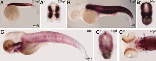
Expression of myl1 in post-somitogenic stages. A: Expression of myl1 was restricted to myogenic structures in the dorsal and ventral part of the trunk in the 24-hpf embryo. A2: Transverse section of 24-hpf embryo showing myl1 expression in the muscle domain. B: Expression of myl1 in 2-dpf embryo in whole mount showing myl1 specifically in the dorsal and ventral trunk. B2: Transverse section of 2-dpf embryo showing myl1 in the myogenic region of the trunk. C: Myl1 expression is expressed in the trunk musculature in 4-dpf juvenile fish, but at decreasing levels as compared to earlier stages of development. C2: Transverse section of 4-dpf fish showing myl1 in the myogenic region of the trunk. C3: Transverse section of 4-dpf fish showing myl1 expression in the craniofacial musculature. |
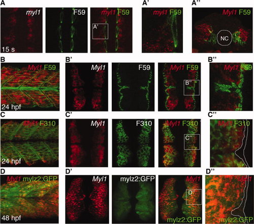
Fibre-type specific expression of myl1. A: Expression of myl1 (red) was detected in the fast domain of the paraxial mesoderm in a 15-somite-stage flat-mounted embryo using fluorescent in situ hybridisation. Labelling of adaxial cells/slow fibres using F59 antibody (green) showed no overlap with the myl1 staining. A2: Blow-up of a single somite as indicated by square in A. A3: Transverse section of 15-somite-stage embryo. NC, notochord. Labelling of adaxial cells/slow fibres using F59 antibody (green) showed no overlap with the myl1 staining. B: Lateral view of 24-hpf embryo stained with F59 antibody (green), labelling slow fibers and myl1 antisense probe (red). B2: Transverse sections of 24-hpf embryo where slow fibres, detected with F59 antibody (green), showed no co-localisation with myl1 antisense probe (red). B3: Blow-up of region boxed in B2. C: Lateral view of 24-hpf embryo where F310 antibody (green) labelling fast fibres is detected in myl1-positive fibres (red). C2: Transverse sections of 24-hpf embryo where fast fibres detected with F310 antibody (green) co-localised with myl1 antisense probe (red). C3: Blow-up of region boxed in C2. D: Lateral view of 48-hpf Tg(mylz2:GFP)i135 transgenic embryo where GFP+ fast fibres were co-labelled with myl1 antisense probe (red). D2: Transverse sections of 48-hpf Tg(mylz2:GFP)i135 transgenic embryo where GFP+ fast fibres were co-labelled with myl1 antisense probe (red). D3: Blow-up of region boxed in D2. |
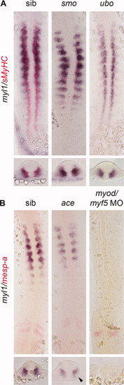
Expression of myl1 at 15-somite stage. myl1 (blue) expression is detected in the fast-twitch muscle domain of the wt embryos as well as of the smo mutant. In the Prdm1 mutant (ubo), myl1 is ectopically expressed in the adaxial cells. smyhc1 (red) labels adaxial cells. B: In FGF8a mutant (ace), myl1 (blue) expression is reduced due to loss of the lateral part of the fast domain (arrowhead in the transversal section). In MyoD/Myf5 morphantsthere is a depletion of myl1 expression. mesp-a labels somite primordia. |
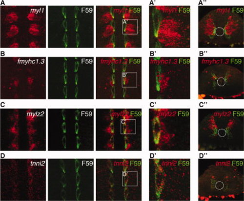
myl1 is the only skeletal fast muscle marker restricted to the skeletal fast muscle domain at 12-somite stage. A: Expression of myl1 (red) is excluded from F59-positive adaxial cells (green) and is specific to the fast domain at 12-somite stage. A2: Blow-up of region boxed in A. A3: Transverse section of 12-somite-stage embryo. B: fmyhc1.3 (red) is co-expressed with F59-positive adaxial cells (green) and is non-specific to the fast domain at 12-somite stage. B2: Blow-up of region boxed in B. B3: Transverse section of 12-somite-stage embryo. C: mylz2 (red) is co-expressed with F59-positive adaxial cells (green) and is non-specific to the fast domain at 12-somite stage. C2: Blow-up of region boxed in C. C3: Transverse section of 12-somite-stage embryo. D: tnni2 (red) is co-expressed with F59-positive adaxial cells (green) and is non-specific to the fast domain at 12-somite stage. D2: Blow-up of region boxed in D. D3: Transverse section of 12-somite-stage embryo. |