
- Title
-
Somatic mutagenesis with a sleeping beauty transposon system leads to solid tumor formation in zebrafish
- Authors
- McGrail, M., Hatler, J.M., Kuang, X., Liao, H.K., Nannapaneni, K., Noack Watt, K.E., Uhl, J.D., Largaespada, D.A., Vollbrecht, E., Scheetz, T.E., Dupuy, A.J., Hostetter, J.M., and Essner, J.J.
- Source
- Full text @ PLoS One
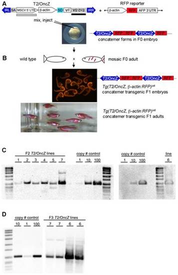
Isolation of transgenic T2/OncZ concatemer lines. (A) T2/OncZ transposon vector and RFP reporter used to isolate concatemers. IRL and IRR, left and right transposon inverted repeats; SA, splice acceptor from intron 1 of ß-actin gene; MSCV 52 LTR, murine stem cell virus 52 long terminal repeat; β-actin, β-actin promoter minus splice acceptor at 3′ end; SD, splice donor; En2-SA, splice acceptor from mouse engrailed 2 gene; pA, SV40 polyadenylation sequence; AFP, ocean pout antifreeze protein 3′ UTR; black bar represents probe used on genomic Southerns shown in panel C. Grey box represents probe used on genomic Southerns shown in panel D. Linear DNA fragments of T2/OncZ and the β-actin:RFP reporter gene were mixed and co-injected into 1-cell zebrafish embryos. (B) Adult F0 founders were outcrossed to wild type and transgenic F1 embryos identified by ubiquitious RFP fluorescence. (C) Genomic Southern blots to estimate transposon copy number in Tg(T2/OncZ, β-actin:RFP) concatemer lines 1–7. DNA was isolated from F2 generation heterozygous adults. (D) Genomic Southern blot of DNA isolated from F3 generation heterozygous Tg(T2/OncZ, β-actin:RFP)is7 and Tg(T2/OncZ, β-actin:RFP)is6 adults. Plasmid pT2/OncZ was loaded as reference in copy # control lanes. |
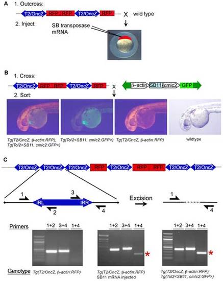
Two strategies for T2/OncZ insertional mutagenesis in zebrafish somatic tissues. (A) Methods 1: Injection of in vitro transcribed SB11 mRNA at the 1-cell stage. (B) Method 2: Genetic cross between Tg(T2/OncZ, β-actin:RFP) (T2/OncZ) and constitutive Tg(Tol2<β-actin:SB11, cmlc2:GFP>) (β-actin:SB11) fish. At 24 hpf embryos are sorted into 4 progeny classes. (C) Excision PCR assay on 5 dpf larvae to demonstrate mobilization of transposons out of the concatemer in the presence of SB11 transposase. Primers 1 and 4 amplify a 220 bp band (red asterisk) flanking the transposon excision site in the concatemer. Left panel, Tg(T2/OncZ, β-actin:RFP)is6 larvae; middle panel, SB11 injected Tg(T2/OncZ, β-actin:RFP)is6 larvae; right panel, double transgenic Tg(T2/OncZ, β-actin:RFP)is6; Tg(Tol2<β-actin:SB11, cmlc2:GFP>) larvae. |
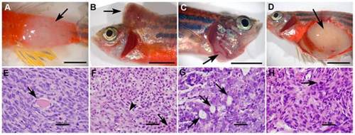
Histopathologic features of solid tumors in T2/OncZ mutagenized fish. (A–D) gross images of neoplasms in fish 1a, 2, 6, and 8 respectively. (E–H) Histopathology of zebrafish neoplasms: Hematoxylin and Eosin stained sections at 1000× magnification. (E) Spindle cell sarcoma from fish 1a, note entrapped skeletal muscle fibers (arrow). (F) Mixed neoplasm from fish 2, neoplastic round cells (arrow) were intermixed with neoplastic spindle shaped cells (arrow head). (G) Carcinoma from fish 6, neoplastic cells were arranged into multiple acinar structures (arrow). (H) Spindle cell sarcoma from fish 8, note the mitotic figure (arrow). Scale bar A–D, 0.5 cm; E–H, 50 μm. |