
|
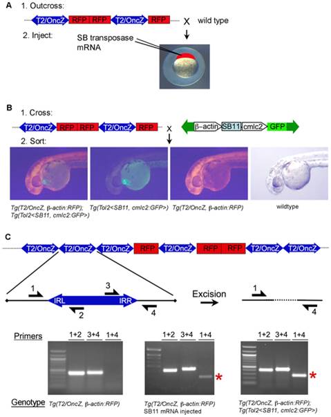
Fig. 3 Two strategies for T2/OncZ insertional mutagenesis in zebrafish somatic tissues.
(A) Methods 1: Injection of in vitro transcribed SB11 mRNA at the 1-cell stage. (B) Method 2: Genetic cross between Tg(T2/OncZ, β-actin:RFP) (T2/OncZ) and constitutive Tg(Tol2<β-actin:SB11, cmlc2:GFP>) (β-actin:SB11) fish. At 24 hpf embryos are sorted into 4 progeny classes. (C) Excision PCR assay on 5 dpf larvae to demonstrate mobilization of transposons out of the concatemer in the presence of SB11 transposase. Primers 1 and 4 amplify a 220 bp band (red asterisk) flanking the transposon excision site in the concatemer. Left panel, Tg(T2/OncZ, β-actin:RFP)is6 larvae; middle panel, SB11 injected Tg(T2/OncZ, β-actin:RFP)is6 larvae; right panel, double transgenic Tg(T2/OncZ, β-actin:RFP)is6; Tg(Tol2<β-actin:SB11, cmlc2:GFP>) larvae.