
- Title
-
Distinct and Conserved Prominin-1/CD133-Positive Retinal Cell Populations Identified across Species
- Authors
- Jászai, J., Fargeas, C.A., Graupner, S., Tanaka, E.M., Brand, M., Huttner, W.B., and Corbeil, D.
- Source
- Full text @ PLoS One
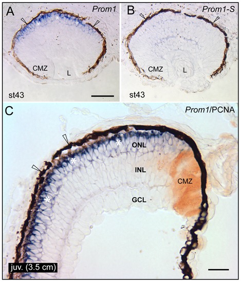
Localization of prominin-1 in the retina of axolotl. (A–C) Cryosections of eyes from larval (A, B; stage 43) and juvenile (C; juv, length of 3.5 cm) axolotls were processed for non-radioactive ISH using either antisense (A, C; Prom1) or sense (B; Prom1-S) DIG-labelled prominin-1 probe alone (A, B) or combined with IHC detection of proliferating cell nuclear antigen (C; PCNA). Asterisks indicate prominin-1–positive cell bodies (A, C, blue) located within the outer nuclear layer (ONL). Arrowheads indicate the retinal pigmented (dark brown) epithelium. Note the lack of prominin-1 reactivity within the ciliary marginal zone (CMZ), which is labelled with PCNA (C, brown). L, lens; INL, inner nuclear layer; GCL; ganglion cell layer. Scale bars, A and B, 100 μm; C, 50 μm. |
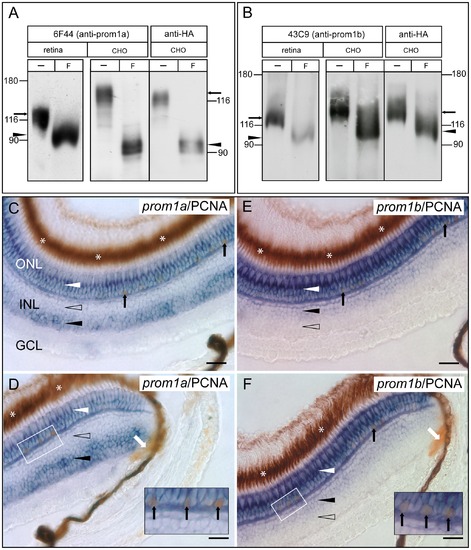
Expression and differential distribution of prominin-1a and b in the retina of zebrafish. (A, B) Lysates prepared from either zebrafish retina (retina) or CHO cells transiently transfected with either Dr prominin-1a (A, CHO) or prominin-1b. (B, CHO) both fused in-frame to a C-terminal HA-epitope tag were incubated in absence (-) or presence (F) of PNGase F and analyzed by immunoblotting with either rabbit antiserum 6F44 (6F44; anti-prominin-1a) or 43C9 (43C9; anti-prominin-1b) or rat mAb anti-HA (anti-HA). Arrow and arrowhead indicate native and N-deglycosylated forms of Dr prominin-1a (A) or prominin-1b (B), respectively. The position of prestained apparent molecular mass markers (in kDa) is indicated. (C–F) Cryosections of eyes from adult zebrafish were processed for non-radioactive ISH using antisense DIG-labelled prominin-1a (C, D; prom1a) or prominin-1b (E, F; prom1b) probe combined with IHC detection of proliferating cell nuclear antigen (PCNA). Central (C, E) and peripheral (D, F) regions are presented. White arrowheads indicate prominin-1a– and prominin-1b–positive cell bodies (blue) located in the outer nuclear layer (ONL). Black and hollow arrowheads indicate the presence or absence of a given prominin-1 molecule reactivity within the inner nuclear layer (INL), respectively. Black and white arrows point to PCNA–positive nuclei (brown) within the ONL and ciliary marginal zone, respectively. Asterisks indicate the retinal pigmented (dark brown) epithelium. The boxed area demarcates a region displayed at higher magnification in the inset of the corresponding panel, where scarce prominin-1/PCNA–positive cells are observed. Notice that, in addition to the overlapping signals of both zebrafish co-orthologues of mammalian prominin-1 in the ONL, a differential expression could be observed within the INL with respect to the scleral and vitreal subdivisions. GCL, ganglion cell layer. Scale bars, 25 μm. EXPRESSION / LABELING:
|
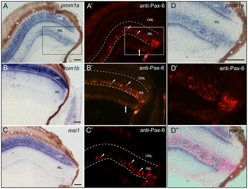
Co-expression analysis of zebrafish prominin-1a or b with Pax-6 and/or musashi-1 within retina cell layers. (A–D′′) Cryosections of eyes from adult zebrafish were processed for non-radioactive ISH using antisense DIG-labelled prominin-1a (A, D, D′′; prom1a), prominin-1b (B; prom1b) or musashi-1 (C; msi1) probe combined with IHC detection of Pax-6 (A′–C′, D′, D′′; anti-Pax-6). Arrowheads and arrows show the Pax-6 immunoreactivity (red) within the inner nuclear layer (INL) and ganglion cell layer (GCL), respectively. The area shown in the inset (A, A′) is displayed as zoom-in in panels D-D3. Note the expression of Pax-6 overlaps with prominin-1a within the INL. Asterisks indicate the retinal pigmented (dark brown) epithelium. Dashed lines delineate the INL. e, erythrocytes. Scale bars, 25 μm. EXPRESSION / LABELING:
|