
|
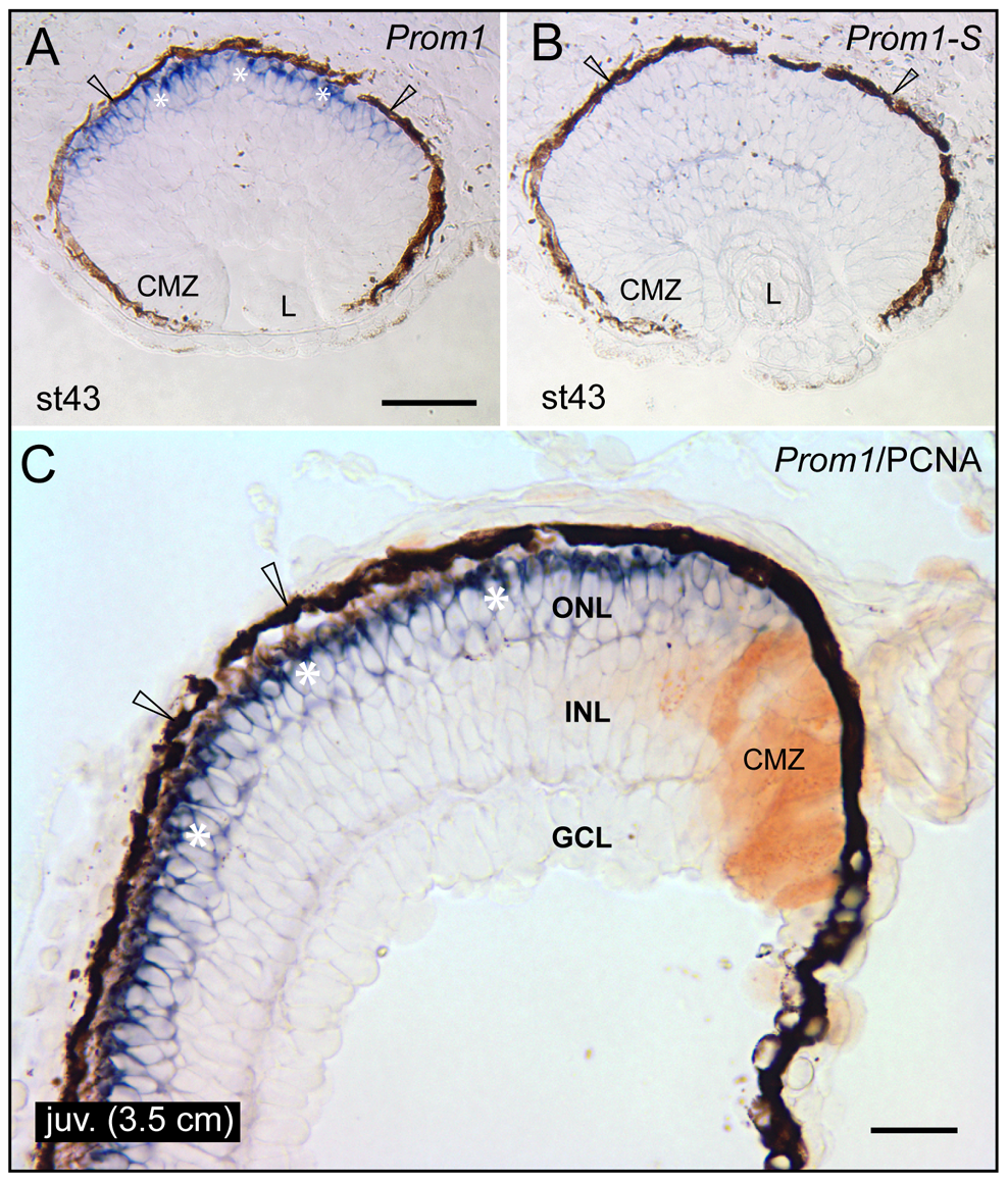
Fig. 1
Localization of prominin-1 in the retina of axolotl.
(A–C) Cryosections of eyes from larval (A, B; stage 43) and juvenile (C; juv, length of 3.5 cm) axolotls were processed for non-radioactive ISH using either antisense (A, C; Prom1) or sense (B; Prom1-S) DIG-labelled prominin-1 probe alone (A, B) or combined with IHC detection of proliferating cell nuclear antigen (C; PCNA). Asterisks indicate prominin-1–positive cell bodies (A, C, blue) located within the outer nuclear layer (ONL). Arrowheads indicate the retinal pigmented (dark brown) epithelium. Note the lack of prominin-1 reactivity within the ciliary marginal zone (CMZ), which is labelled with PCNA (C, brown). L, lens; INL, inner nuclear layer; GCL; ganglion cell layer. Scale bars, A and B, 100 μm; C, 50 μm.