
- Title
-
Zebrafish embryo screen for mycobacterial genes involved in the initiation of granuloma formation reveals a newly identified ESX-1 component
- Authors
- Stoop, E.J., Schipper, T., Rosendahl Huber, S.K., Nezhinsky, A.E., Verbeek, F.J., Gurcha, S.S., Besra, G.S., Vandenbroucke-Grauls, C.M., Bitter, W., and van der Sar, A.M.
- Source
- Full text @ Dis. Model. Mech.
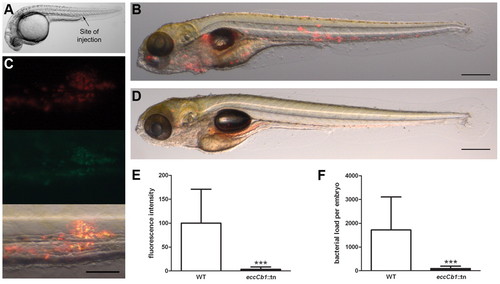
Initiation of granuloma formation in M. marinum E11 (Mma11)-infected embryos. (A) Overview of a zebrafish embryo at 28 hpf. The arrow indicates the caudal vein injection site used in this study. (B) Embryo 5 days after infection with 110 CFUs Mma11. Overlay of brightfield and fluorescent images is shown. Aggregates of red fluorescent bacteria are seen in the tail and head region. Scale bar: 500 μm. (C) Bacterial clustering in tail at 5 dpi with 92 CFUs Mma11 expressing hsp::DsRed and gap7::eGFP. Of the total amount of bacteria [constitutively expressing red fluorescence (top panel)], the majority expresses eGFP (middle panel), indicating that the bacteria reside in clusters that resemble granulomas. Overlay shows overlapping red and green fluorescence as yellow (bottom panel). Scale bar: 50 μm. (D) The Mma11 eccCb1::tn mutant is highly attenuated for initiation of granuloma formation at 5 dpi with 171 CFUs. Loose spots of red fluorescent bacteria are detected, but aggregates are not found. Scale bar: 500 μm. (E) Quantification of infection as determined with specially designed software. The amount of red (fluorescent) pixels of fluorescent images of embryos infected with the eccCb1::tn mutant is set as a percentage of the amount of red pixels of fluorescent images of embryos infected with wild-type bacteria (WT). Data shown are mean + standard deviation of three independent experiments (***P<0.001, unpaired Student’s t-test). (F) From embryos used in E, bacterial loads were determined by plating whole embryos (***P<0.001, unpaired Student’s t-test). |
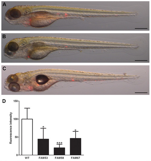
Three out of 200 transposon mutants were identified as early granuloma mutants. Infection of embryos with mutants FAM53, FAM58 and FAM67 reproducibly resulted in either smaller or fewer early granulomas at 5 dpi, as compared with the wild type (see Fig. 1B). (A–C) Representative overlays of brightfield and fluorescent images of embryos infected with 71 CFUs of mutant FAM53 (A), 70 CFUs of mutant FAM58 (B) and 86 CFUs of mutant FAM67 (C) are shown. Scale bars: 500 μm. (D) Relative levels of infection determined by automated quantification of fluorescent pixels. The results represent mean + standard deviation of all experiments performed (n=3 to 9) (*P<0.029, ***P<0.001, unpaired Student’s t-test). |
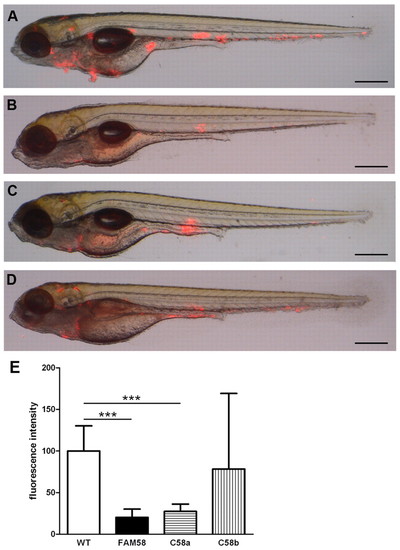
Complementation of early granuloma mutant FAM58. Embryos 5 days after infection with wild-type Mma11, mutant FAM58 and FAM58 carrying a plasmid for expression of espL (C58a) or the espL-espB operon (C58b). (A–D) Representative images of embryos for all experiments performed (n=4 to 9) are shown. Infection doses were 137 CFUs for wild-type Mma11 (A), 128 CFUs for mutant FAM58 (B), 171 CFUs for C58a (C) and 127 CFUs for C58b (D). Scale bars: 500 μm. (E) Automated quantification of relative infection level. Results represent mean + standard deviation for all experiments performed (n=4 to 9) (***P<0.001, unpaired Student’s t-test). |