FIGURE SUMMARY
Title

The Leukemia-Associated Mllt10/Af10-Dot1l Are Tcf4/{beta}-Catenin Coactivators Essential for Intestinal Homeostasis

Authors
Mahmoudi, T., Boj, S.F., Hatzis, P., Li, V.S., Taouatas, N., Vries, R.G., Teunissen, H., Begthel, H., Korving, J., Mohammed, S., Heck, A.J., and Clevers, H.
Source
Full text @ PLoS Biol.

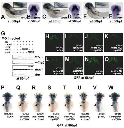
tcf7l2, Mllt10/Af10, and dot1l are co-expressed in Wnt active tissues and required for TCF-driven transcription in vivo.
(A–F) In situ hybridization for tcf7l2 (A), mllt10/af10 (C), and dot1l (E) in whole embryos at 80 hpf. In situ hybridization for tcf7l2 (B), mllt10/af10 (D), and dot1l (F) in whole embryos at 56 hpf. Arrowhead indicates rhombomeres. (G) Gene-specific depletion of RNA levels at 80 hpf determined by RT-PCR analysis of tcf7l2, mllt10, and dot1l expression in whole embryos injected with the indicated MO sequences. Higher bands in the RTPCR panels for tcf7l2 and dot1l correspond to unspliced introns in the PCR products. Morpholino knockdown of mllt10/af10 and dot1l mimics tcf7l2 depletion effect on Wnt reporter activity in zebrafish brain (H–M). Representative confocal images of dorsal view of head of 56 hpf TOPdGFP embryos injected with different MOs. (H) dGFP expression in the hindbrain of embryos injected with buffer only, MO against tcf7l2 (I), two independent mllt10/af10 (J,K) and dot1l MOs (L,M), p53 MO (N), and control MO (O). Depletion of mllt10/af10 and dot1l abrogate GFP expression in intestine of TOPdGFP zebrafish, mimicking tcf7l2 depletion. Representative dorsal view of whole mount in situ hybridization for GFP in heterozygous TOPdGFP embryos at 80 hpf injected with (P) buffer alone, MOs against tcf7l2 (Q), mllt10/af10 (R,S), dot1l (T,U), p53 (V) and control MO (W). All MOs have been coinjected with a MO against p53.

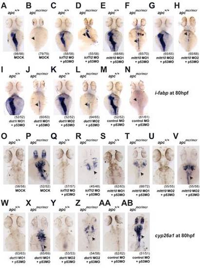
Depletion of mllt10/af10 and dot1l rescues intestinal defects in apcmcr/mcr zebrafish, mimicking tcf7l2 depletion.
Depletion of mllt10/af10 and dot1l rescues expression of i-fabp in apcmcr/mcr mutant embryos, placing these genes downstream of Apc as Wnt target gene activators (A–N). Representative whole mount in situ hybridizations for i-fabp in wild type and apcmcr/mcr mutant embryos at 80 hpf injected with (A,B) buffer alone,(C,D) MO against tcf7l2, (E–H) two independent mllt10/af10 MOs, (I–L) two independent dot1l MO, and (M,N) control MO. Depletion of mllt10/af10 and dot1l rescues mis-expression of cyp26a1 in apcmcr/mcr mutant embryos (O–AB). Representative whole mount in situ hybridizations for cyp26a1 in wild type and apcmcr/mcr mutant embryos at 80 hpf injected with (O,P) buffer alone, (Q,R) MO against tcf7l2, (S–V) two independent mllt10/af10 MOs, (W–Z) two independent dot1l MO, and (AA,AB) control MO. All MOs have been coinjected with an MO against p53. All images were captured using the same exposure and represent at least three independent experiments. In parentheses number of embryos showing described phenotype per number of total embryos analyzed.

tcf7l2, mllt10/af10, and dot1l are essential for Wnt target gene expression and intestinal homeostasis in zebrafish.
(A–H) Representative image of BrdU incorporation into epithelial cells in the intestinal bulb of 124 hpf zebrafish embryos. BrdU staining in wild-type embryos (A) and embryos depleted of tcf7l2 (B), mllt10/af10 (C,D), and dot1l (E,F), p53 (G), and control (H) by MO injection. (I) Quantification (mean ± SD) of percent BrdU positive cells and (J) total number of cells in the epithelial layer of the intestinal bulb (20 embryos/condition). * p<0.05. Depletion of tcf7l2, mllt10/af10, and dot1l MO abrogates axin2 expression in intestine. In situ hybridization for axin2 in 80 hpf wild-type and MO-injected embryos (K–R). Staining for axin2 in wild type embryos uninjected (K) or injected with MO against tcf7l2 (L), mllt10/af10 (M,N), and dot1l (O,P), p53 (Q), and control MO (R). All MOs have been coinjected with a MO against p53. In parentheses number of embryos with phenotype per total analyzed. Images are representative of three independent experiments. sb, swim bladder; ib, intestinal bulb. Scale bar: 100 μm.

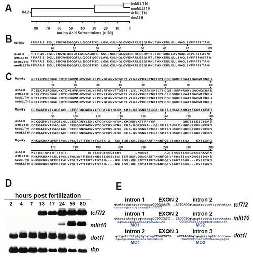
mRNA expression of tcf7l2, mllt10, and dot1l during zebrafish embryonic development and their depletion by splice-inhibiting MO sequences. (A) Phylogenetic tree analysis places zebrafish (Danio rerio) Mllt10 close to mouse and human sequences. (B–C) Amino acid sequence alignment of the Leucine zipper and PHD finger domains show high conservation of Mllt10 between species. (D) RT-PCR analysis of tcf7l2, mllt10, dot1l, and tbp RNA expression levels in whole embryos at different stages of embryonic development. (E) Representation of the splice-blocking MO sequences (blue) generated to deplete mRNA levels for tcf7l2, mllt10, and dot1l.

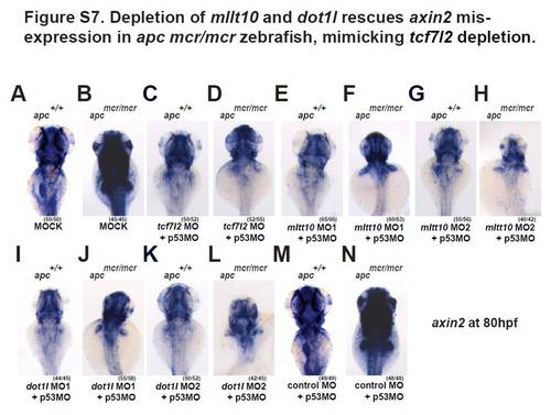
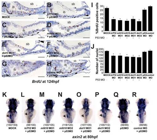
Depletion of tcf7l2, mllt10/af10 and dot1l rescues mis-expression of axin2 in apcmcr/mcr zebrafish, placing these genes downstream of Apc as Wnt target gene activators (A–N). Representative whole mount in situ hybridizations for axin2 in wild type and apcmcr/mcr mutant embryos at 80 hpf injected with (A,B) buffer alone, (C,D) MO against tcf7l2, (E–H) two independent mllt10/af10 MOs, (I–L) two independent dot1l MO, and (M,N) control MO. All MOs have been coinjected with a MO against p53. All images were captured using the same exposure and represent at least three independent experiments. In parentheses number of embryos showing described phenotype per number of total embryos analyzed.

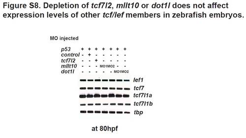
Depletion of tcf7l2, mllt10, or dot1l does not affect the expression levels of other tcf/lef family members in zebrafish embryos. RT-PCR analysis of lef1, tcf7, tcf7l1a, tcf7l1b, and tbp RNA expression levels in whole embryos injected with MO against p53 alone, or MO against p53 coinjected with MOs against tcf7l2, mllt10, dot1l, or control MO, respectively.

Acknowledgments
This image is the copyrighted work of the attributed author or publisher, and ZFIN has permission only to display this image to its users. Additional permissions should be obtained from the applicable author or publisher of the image. Full text @ PLoS Biol.