
- Title
-
A forward chemical screen in zebrafish identifies a retinoic acid derivative with receptor specificity
- Authors
- Das, B.C., McCartin, K., Liu, T.C., Peterson, R.T., and Evans, T.
- Source
- Full text @ PLoS One
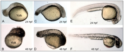
BT10 added during early embryogenesis causes a major disruption of the body axis. Shown are representative embryos (from 3 experiments, for each sample n = 20) treated with BT10 at 10 μM (A-D) compared to embryos treated only with vehicle (DMSO) as control. BT10 was added at 5 hpf (A, B) or 8 hpf (C, D). DMSO alone was added at 5 hpf (E, F). Embryos were allowed to develop until 24 hpf (A, C, E) or 48 hpf (B, D, F). The position of the yolk is indicated in panels A and B. All embryos are viewed laterally, anterior to the right, dorsal at the top. |

BT10 causes specific cardiovascular defects when added at 24 hpf. Shown are representative embryos (from 3 experiments, for each sample n = 20) that were cultured between 24–60 hpf in the presence of DMSO alone (A, control) or BT10 (B). Note the defined morphological structure of the ventral yolk sac (arrow). A normal looping heart tube fails to develop in these embryos. Panels C and D show higher magnification views of representative control (C) and BT10-treated (D) embryos, respectively. The red block arrow indicates blood flowing through the normal control heart, which is not evident in the BT10-treated embryos. Views are lateral, anterior to the right, dorsal at the top. PHENOTYPE:
|

BT10 added at 24 hpf causes a defect in heart tube looping, similar to excess RA. Shown are representative embryos (n = 30) containing a cmlc2:gfp transgene were cultured in vehicle alone (control) or in the presence of ATRA or BT10 at the concentrations indicated, and as described in Fig. 3B, beginning at 24 hpf. They were then photographed at 48 hpf in both brightfield and fluorescence, and the images overlayed to document the morphology of the heart tube (green). In control embryos the ventricle (V) is fully looped relative to the atrium (A), indicated by the dotted white line. In contrast, embryos cultured in the presence of ATRA or BT10 develop a relatively linear heart tube (at “low” or “high” concentrations), which is the major obvious morphological abnormality. |
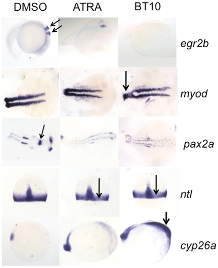
Changes in the expression patterns for known retinoid target genes confirm that BT10 is a retinoid agonist. Shown are representative embryos (n = 30) treated with vehicle (DMSO, left panels), 0.1 μM ATRA (middle panels), or 5 μM BT10 (right panels) and then analyzed by in situ hybridization at the 10 somite stage (cyp26a), 75% epiboly (ntl), the 18 somite stage (myod and pax2a), or the 26 somite stage (egr2b). Embryos are viewed laterally with anterior to the right (egr2b, cyp26a), dorsally with anterior to the right (myod, pax2a), or dorsally with animal pole to the top (ntl, arrows in both panels). Note that in both ATRA and BT10 treated embryos, the normal anterior expression domains are largely abolished (egr2b, arrows indicate normal expression pattern marking the 3rd and 5th rhombomeres in the hindbrain of control embryos, and pax2a, arrow indicates normal expression pattern in the midbrain-hindbrain boundary domain), highly reduced (myod, disruption noted by arrow in BT10 panel), or more subtly inhibited (ntl, indicated by arrows in both panels). In contrast, the expression domain of cyp26a is expanded in BT10-treated embryos throughout the anterior region (arrow in BT10 panel). EXPRESSION / LABELING:
|