
- Title
-
Zebrafish have a competent p53-dependent nucleotide excision repair pathway to resolve ultraviolet B-induced DNA damage in the skin
- Authors
- Zeng, Z., Richardson, J., Verduzco, D., Mitchell, D.L., and Patton, E.E.
- Source
- Full text @ Zebrafish
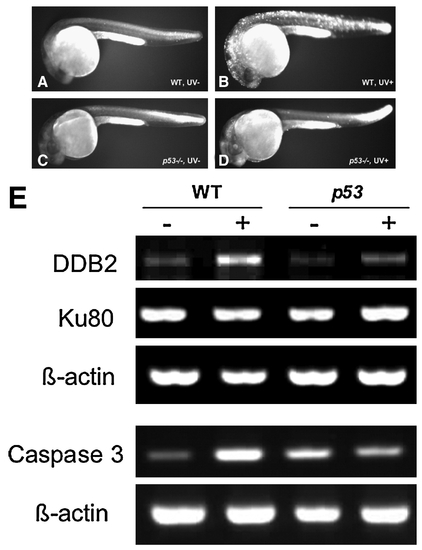
UVB-induced apoptosis and expression of DNA damage repair–related genes. (A–D) Twenty-four hpf wt and p53 mutant embryos were irradiated with a sublethal dose of UVB (1.08 kJ/m2). After incubated in dark for 6h, embryos were stained with acridine orange solution to detect cell death induced by UVB treatment in living embryos. (E) Total RNA isolated and reverse transcription polymerase chain reaction analysis was performed to examine the expression of DNA damage repair–related genes after UVB exposure. wt, wild type. |
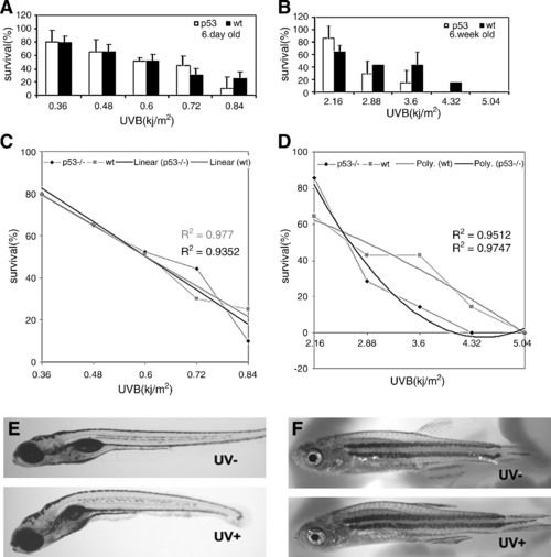
p53 Mutant zebrafish are sensitive to UVB irradiation. (A) Comparison of sensitivity of 6-day-old wt and p53 mutant fish at various UVB doses. Ten fish were used at each dose in each experiment. Experiments had three replicates. Means and standard deviations of the three experiments are presented. (B) Comparison of sensitivity in 5–6-week-old wt and p53 fish at various UVB doses. (C) The data from 6-day-old wt and p53 fish fit to linear regression lines (R2 = 0.977 and 0.9352 for wt and p53 fish, respectively; R2 = 1 when curves best fit the regression line). The calculated LD50s (where survival is 50%) are 0.6 kJ/m2 for both wt and p53 fish. (D) The data from 5–6-week-old wt and p53 young adults fit to polynomial regression lines (R2 = 0.9512 and 0.9747 for wt and p53 fish, respectively). (E, F) The typical morphology of UVB-exposed zebrafish. Curved spinal is the common abnormalities in both juvenile and young adult fish, also in wt and p53 mutant fish. PHENOTYPE:
|
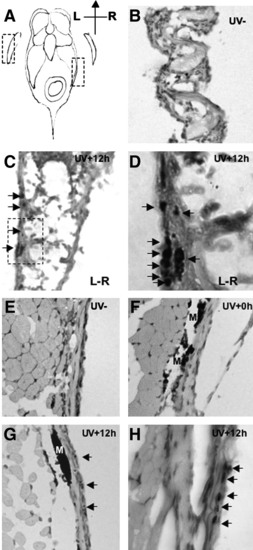
Observation of DNA damage sites in zebrafish fin (B–D) and skin (E–H). Histological sections of adult zebrafish 12 hours after of UVB (2.16kJ/m2) exposure, stained with an antibody to detect phospho-histone H2AX (phospho-H2AX). (A) Schematic diagram of zebrafish section; dotted boxes denote the examined fin and skin tissues in (B) and (E), respectively. (B) Section of pectoral fin of untreated wt fish (hematoxylin and eosin stain; magnification 200x). (C) Section of pectoral fin of UVB exposed wt fish. Phospho-H2AX signals are located in the epidermis on the most outer side of fin (arrows); magnification 200x. Dotted box indicates region examined in (D). (D) High magnification (630x) view of the phospho-H2AX positive cells in the fin. (E) No phospho-H2AX signal is detected in the skin of untreated fish; magnification 200x. (F) Skin of fish sacrificed immediately after UVB exposure reveals no staining for phospho-H2AX; magnification 200x. (G) Skin at 12 hours postirradiation reveals specific phospho-H2AX staining; magnification 200x. (H) High magnification (630x) view of the phospho-H2AX positive cells in the skin. Phospho-H2AX positive nuclei are indicated with arrows; M denotes melanin. |

ZFIN is incorporating published figure images and captions as part of an ongoing project. Figures from some publications have not yet been curated, or are not available for display because of copyright restrictions. PHENOTYPE:
|