
|
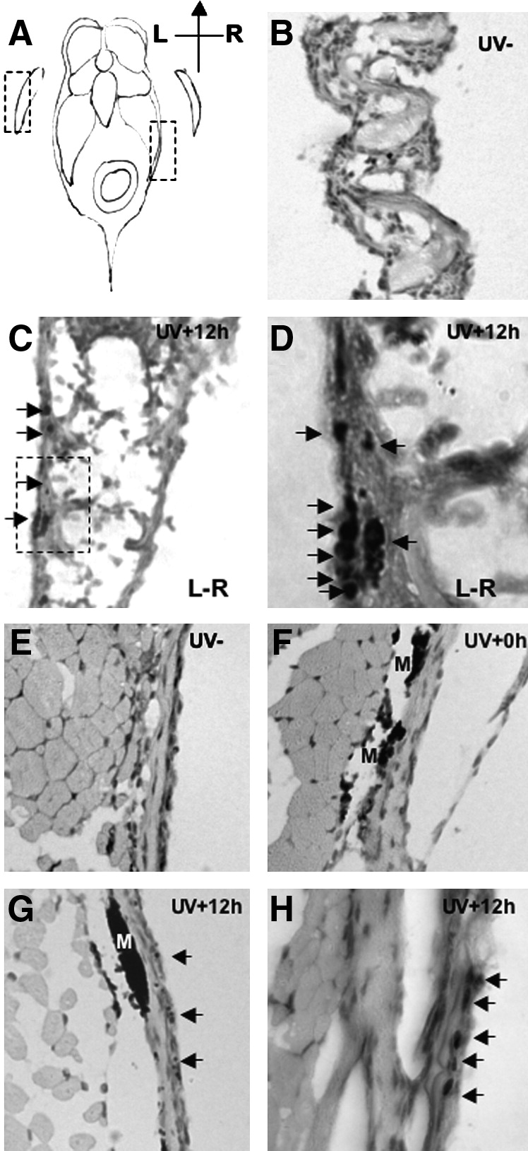
Fig. 4 Observation of DNA damage sites in zebrafish fin (B–D) and skin (E–H). Histological sections of adult zebrafish 12 hours after of UVB (2.16kJ/m2) exposure, stained with an antibody to detect phospho-histone H2AX (phospho-H2AX). (A) Schematic diagram of zebrafish section; dotted boxes denote the examined fin and skin tissues in (B) and (E), respectively. (B) Section of pectoral fin of untreated wt fish (hematoxylin and eosin stain; magnification 200x). (C) Section of pectoral fin of UVB exposed wt fish. Phospho-H2AX signals are located in the epidermis on the most outer side of fin (arrows); magnification 200x. Dotted box indicates region examined in (D). (D) High magnification (630x) view of the phospho-H2AX positive cells in the fin. (E) No phospho-H2AX signal is detected in the skin of untreated fish; magnification 200x. (F) Skin of fish sacrificed immediately after UVB exposure reveals no staining for phospho-H2AX; magnification 200x. (G) Skin at 12 hours postirradiation reveals specific phospho-H2AX staining; magnification 200x. (H) High magnification (630x) view of the phospho-H2AX positive cells in the skin. Phospho-H2AX positive nuclei are indicated with arrows; M denotes melanin.