
- Title
-
The tight junction component Claudin E is required for zebrafish epiboly
- Authors
- Siddiqui, M., Sheikh, H., Tran, C., and Bruce, A.E.
- Source
- Full text @ Dev. Dyn.

Whole-mount in situ hybridization analysis of zebrafish cldne. All embryos shown in lateral view, except (D) which is an animal pole view. A: Ubiquitous maternal expression of cldne at the eight-cell stage. B: Arrowhead indicates patchy cldne expression in the enveloping layer (EVL) at dome stage. C,D: Expression of cldne throughout the EVL at shield stage. E: Arrow indicates more intense hybridization in the EVL at the dorsal margin at 80% epiboly stage. Inset shows flat-mounted dorsal margin of an embryo stained for cldne (purple) and the dorsal forerunner marker cas (red). F: A 1 day postfertilization (dpf) embryo, in addition to faint cldne expression throughout the skin, cldne is detected in the olfactory placodes (arrow), ears (arrowhead), and proctodeum (asterisk). |

Embryos injected with cldne-morpholino oligonucleotide (MO) exhibit epiboly delays. Lateral views of live embryos, injected construct indicated in lower right. A,B: Uninjected embryo at 30% epiboly (A), time matched cldne-MO injected embryo at sphere stage (B). C-E: Uninjected embryo at shield stage (C), time matched cldne-MO injected embryo at 40% epiboly (D), same embryo as in D (E), arrow indicates the yolk syncytial layer (YSL) nuclei, which have compacted normally. F: Shield stage embryo injected with mismatch morpholino. G: Shield stage embryo injected with cldne RNA. H: Rescued embryo injected with cldne RNA and cldne-MO. I,J: Uninjected embryo at 90% epiboly stage (I), time matched cldne-MO injected embryo at 70% epiboly (J). PHENOTYPE:
|
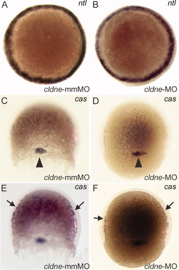
Deep cell fate specification occurs normally in cldne morphant embryos. Probe indicated in upper right, injected construct indicated in lower right. A,B: Animal pole views of shield stage embryos stained for ntl. C-F: Lateral views of 70% epiboly embryos stained for cas. Focal plane in C and D shows dorsal forerunner cells (arrowhead), while the focal plane in E and F shows scattered endodermal cells adjacent to the yolk (arrows). |
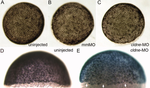
Enveloping layer (EVL) specification is normal in cldne morphant embryos. A-E: Injected constructs indicated in lower right (A-C) and upper right (D,E). A-C: Animal pole views. D,E: Lateral views. E: EVL margin is irregular in cldne-MO injected embryos (arrows). |

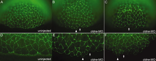
The tight junction associated protein ZO-1 is present in morphant embryos at shield stage. A-D: Uninjected embryos (A,B) and cldne-morpholino oligonucleotide (MO) injected embryos (C,D) stained for ZO-1. A-D show lateral (A,B,D) or animal pole (C) views. B,D: Arrows indicate the EVL margin, with the yolk cell at the bottom. |
Phalloidin staining reveals abnormal marginal enveloping layer (EVL) cell morphology. A-F: Lateral views of phalloidin stained embryos, injected constructs indicated in lower right. Arrowheads indicate abnormally round marginal EVL cells; arrows indicate gaps between cells. A-C: Embryos at dome to 30% epiboly stage. D-F: Embryos at shield stage. PHENOTYPE:
|

Unillustrated author statements PHENOTYPE:
|