
- Title
-
fezf2 expression delineates cells with proliferative potential and expressing markers of neural stem cells in the adult zebrafish brain
- Authors
- Berberoglu, M.A., Dong, Z., Mueller, T., and Guo, S.
- Source
- Full text @ Gene Expr. Patterns
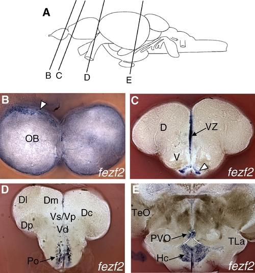
fezf2 mRNA expression in the adult zebrafish brain by in situ hybridization. (A) Schematic of adult zebrafish brain showing plane of section in B–E. (B) Coronal section through caudal olfactory bulb showing fezf2 expression in the periphery (arrowhead) (10x magnification). (C) Coronal section through telencephalon showing strong fezf2 expression in the pallial ventricular zone (midline), as well as bilateral ventral expression (arrowhead) (10x magnification). (D) Coronal section showing fezf2 expression in the preoptic region (10x magnification). (E) Coronal section showing fezf2 expression in the hypothalamus (10x magnification). Schematic modified from Wullimann et al., 1996. Abbreviations: OB, olfactory bulb; D, dorsal telencephalon; V, ventral telencephalon; VZ, ventricular zone; Dl, lateral zone of D; Dm, medial zone of D; Dc, central zone of D; Dp, posterior zone of D; Vs, supracommissural nucleus of V; Vp, postcommissural nucleus of V; Vd, dorsal nucleus of V; Po, preoptic region; TeO, tectum opticum; PVO, paraventricular organ; Hc, caudal hypothalamus; TLa, torus lateralis. EXPRESSION / LABELING:
|
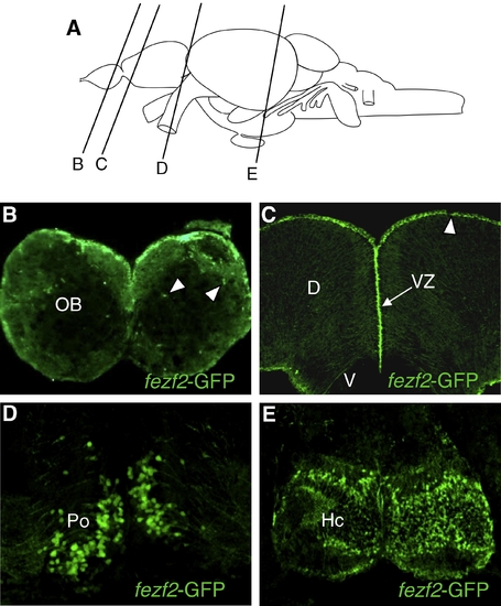
fezf2-GFP transgenic line drives reporter expression in a similar pattern as fezf2 transcripts. (A) Schematic of adult zebrafish brain showing plane of section in B–E. (B) Coronal section through caudal olfactory bulb showing fezf2 expression broadly in the periphery as well as additional fezf2-GFP+ cells in the more internal layers (arrowheads) (20x magnification). (C) Coronal section through telencephalon showing fezf2 expression in the pallial ventricular zone (20x magnification). Note that GFP expression is also observed at the dorsal telencephalic ventricular surface (arrowhead), which is not seen with fezf2 mRNA expression. (D) Coronal section showing fezf2 expression in the preoptic region (40x magnification). (E) Coronal section showing fezf2 expression in the hypothalamus (40x magnification). Schematic modified from Wullimann et al., 1996. Abbreviations: OB, olfactory bulb; D, dorsal telencephalon; V, ventral telencephalon; VZ, ventricular zone; Po, preoptic region; Hc, caudal hypothalamus. EXPRESSION / LABELING:
|
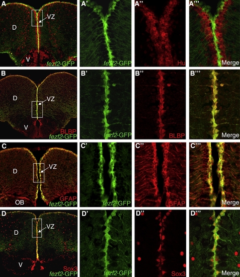
fezf2 is expressed in radial glial cells of the telencephalic ventricular zone, which colocalize with markers of neural stem cells and proliferation. (A) Coronal section through telencephalon showing double-label of fezf2-GFP (green) and Hu (red) (20x magnification). (A′–A′″) Closer view of the boxed region (100x magnification) shows that fezf2-GFP+ cells have radial glial morphology (A′) and do not overlap with neuronal marker HuC/D (A″′). (B) Coronal section through telencephalon showing colocalization of fezf2-GFP (green) and BLBP (red) in the pallial ventricular zone (20x magnification). (B′–B″′) Single confocal Z-section (∼0.5 μm) of the boxed region (100x magnification) shows that fezf2-GFP expression colocalizes precisely with BLBP+ radial glial cells. (C) Coronal section through anterior telencephalon showing double-label of fezf2-GFP (green) and GFAP (red) (20x magnification). (C′–C′″) Single confocal Z-section of the boxed region (40x magnification) shows that fezf2-GFP+ cells colocalize with neural stem cell marker GFAP. (D) Coronal section through telencephalon showing colocalization of fezf2-GFP (green) and Sox3 (red) in the pallial ventricular zone (20x magnification). (D′–D″′) Single confocal Z-section of the boxed region shows that fezf2-GFP+ radial glial cells colocalize with neural stem cell marker Sox3 (100x magnification). EXPRESSION / LABELING:
|
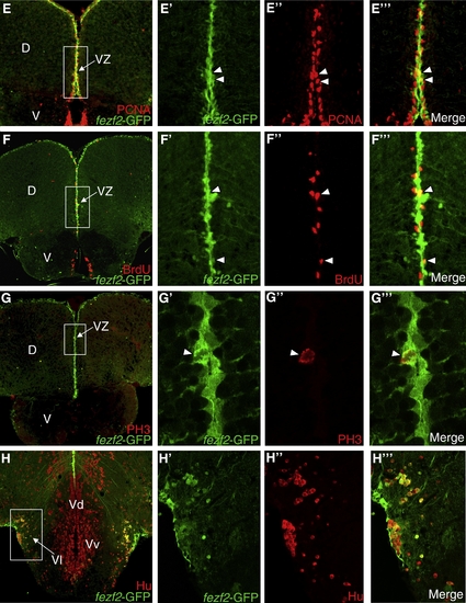
(E) Coronal section through telencephalon showing double-label of fezf2-GFP and proliferation marker PCNA (20x magnification). (E′–E″′) Single confocal Z-section of the boxed region shows that some fezf2-GFP+ cells colocalize with PCNA (arrowheads) (40x magnification). (F) Coronal section through telencephalon showing double-label of fezf2-GFP and BrdU (Bromodeoxyuridine, S-phase marker) (20x magnification). (F′–F″′) Single confocal Z-section of the boxed region shows that some fezf2-GFP+ cells colocalize with BrdU (arrowheads) (40x magnification). (G) Coronal section through telencephalon showing double-label of fezf2-GFP and PH3 (phospho-histone H3; marker of mitosis) (20x magnification). (G′–G″′) Single confocal Z-section of the boxed region shows colocalization of a fezf2-GFP+ cell with PH3 (arrowhead) (100x magnification). (H) Coronal section through telencephalon showing colocalization of fezf2-GFP with Hu (neuronal marker) in the subpallium (Vl region) (20x magnification). (H′–H″′) Single confocal Z-section of the boxed region shows colocalization of fezf2-GFP with Hu in the Vl region (40x magnification). Abbreviations: D, dorsal telencephalon; V, ventral telencephalon; VZ, ventricular zone; OB, olfactory bulb; Vd, dorsal nucleus of V; Vl, lateral nucleus of V; Vv, ventral nucleus of V. EXPRESSION / LABELING:
|
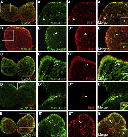
fezf2-GFP+ cells colocalize with markers of neurons and proliferation in the olfactory bulb. (A) Coronal section through the olfactory bulb showing double-labeling of fezf2-GFP (green) and BLBP (red) (20x magnification). (A′–A″′) Some fezf2-GFP+ cells colocalize with neural stem cell/astrocyte marker BLBP (arrowhead), suggesting that these are either local neural stem cells or differentiated astrocytes within the olfactory bulb (40x magnification). (A″′) Inset shows colocalization in a single cell at higher magnification. (B) Coronal section showing fezf2-GFP (green) and GFAP (red) labeling in a single olfactory bulb. (B′–B″′) Some fezf2-GFP+ cells colocalize with GFAP (arrowhead), suggesting that these are either local neural stem cells or differentiated astrocytes (40x magnification). (B″′) Inset shows colocalization in a single cell at higher magnification. (C) Coronal section showing double-labeling of fezf2-GFP (green) and PCNA (red) (20x magnification). (C′–C″′) Some fezf2-GFP+ cells colocalize with proliferation marker PCNA (arrowhead) (40x magnification). (C″′) Inset shows colocalization in a single cell at higher magnification. (D) Coronal section showing double-labeling of fezf2-GFP (green) and BrdU (red) in the caudal olfactory bulb (20x magnification). (D′–D″′) Some medio-dorsal fezf2-GFP+ cells colocalize with BrdU (S-phase marker) (arrowhead) (40x magnification). (D″′) Inset shows colocalization at higher magnification. (E) Coronal section showing double-labeling of fezf2-GFP (green) and Hu (red) (20x magnification). (E′–E″′) Closer view of the boxed region shows that some fezf2-GFP+ cells colocalize with neuronal marker Hu (arrowhead) (40x magnification). (E″′) Inset shows colocalization in a single cell (depicted by arrowhead) at higher magnification. EXPRESSION / LABELING:
|
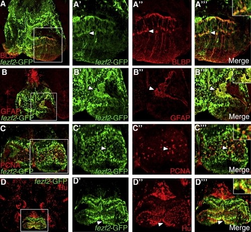
fezf2-GFP is expressed largely in postmitotic neurons of the preoptic region. (A) Coronal section through preoptic region showing double-labeling of fezf2-GFP (green) and BLBP (red) (40x magnification). (A′–A″′) Closer view of the boxed region shows that some fezf2-GFP+ cells colocalize with neural stem cell/astrocytic marker BLBP (arrowhead). (A″′) Inset shows colocalization in a single cell (depicted by arrowhead) at higher magnification. (B) Double-labeling of fezf2-GFP (green) and neural stem cell/astrocytic marker GFAP (red) (40x magnification). (B′–B″′) Closer view of boxed region shows colocalization in some cells (arrowhead). (B″′) Inset shows colocalization in some cells at higher magnification. (C) Double-labeling of fezf2-GFP and proliferating cell marker PCNA (40x magnification). (C′–C″′) Closer view of the boxed region shows that fezf2-GFP+ cells do not co-localize with PCNA. (C″′) Inset shows lack of colocalization at higher magnification. (D) Double-labeling of fezf2-GFP and neuronal marker Hu (40x magnification). (D′–D″′) Closer view of boxed region shows a number of fezf2-GFP+ cells that colocalize with Hu (arrowhead). (D″′) Inset shows colocalization in cells at higher magnification. EXPRESSION / LABELING:
|
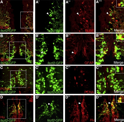
fezf2-GFP expression suggests adult neurogenesis in the caudal hypothalamus. (A) Coronal section through hypothalamus showing double-labeling of fezf2-GFP (green) and BLBP (red) (40x magnification). (A′–A″′) Closer view of the boxed region shows that some fezf2-GFP+ cells colocalize with neural stem cell marker BLBP and have radial glial morphology (arrowhead). (A″′) Inset shows colocalization in cells (depicted by arrowhead) at higher magnification. (B) Double-labeling of fezf2-GFP (green) and neural stem cell marker GFAP (red) (40x magnification). (B′–B″′) Closer view of boxed region shows colocalization in some cells (arrowhead). (B″′) Inset shows colocalization in cells at higher magnification. (C) Double-labeling of fezf2-GFP and proliferating cell marker PCNA (40x magnification). (C′–C″′) Closer view of boxed region shows colocalization in some cells (arrowhead). (C″′) Inset shows colocalization in cells at higher magnification. (D) Double-labeling of fezf2-GFP and neuronal marker Hu (20x magnification). (D′–D″′) Closer view of the boxed region shows colocalization of some fezf2-GFP+ cells with Hu (arrowhead). (D″′) Inset shows colocalization in cells at higher magnification. EXPRESSION / LABELING:
|

Unillustrated author statements EXPRESSION / LABELING:
|
Reprinted from Gene expression patterns : GEP, 9(6), Berberoglu, M.A., Dong, Z., Mueller, T., and Guo, S., fezf2 expression delineates cells with proliferative potential and expressing markers of neural stem cells in the adult zebrafish brain, 411-422, Copyright (2009) with permission from Elsevier. Full text @ Gene Expr. Patterns