
- Title
-
A Systematic Analysis of Tinman Function Reveals Eya and JAK-STAT Signaling as Essential Regulators of Muscle Development
- Authors
- Liu, Y.H., Jakobsen, J.S., Valentin, G., Amarantos, I., Gilmour, D.T., and Furlong, E.E.
- Source
- Full text @ Dev. Cell
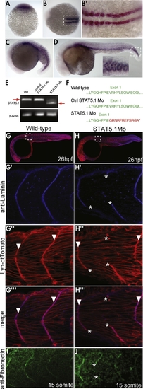
stat5.1 Is Dynamically Expressed during Zebrafish Myogenesis and Is Required for Muscle Morophogenesis (A–D) Whole-mount in situ hybridization of stat5.1 in wild-type embryos at 1-cell (A), 10-somite (B and B′), 15-somite (C), and 24-hpf (D) stages. (B and B′) Dorsal views with anterior to the left. White dashed boxes in (B) and (D) indicate regions with magnified views, shown in (B′) and (D), respectively. (E) RT-PCR analysis of stat5.1 splicing in wild-type, control morpholino, and stat5.1 morpholino-injected embryos. β-actin amplification was used as a control. There is a clear size reduction in stat5.1 morphants due to aberrant splicing in these embryos. The trace amounts of normal spliced transcript visible in the STAT5.1 Mo lane is likely due to variability in embryo injections. (F) Sequencing of uninjected, control morpholino-injected, and stat5.1 morpholino-injected embryos reveals the nature of the aberrant splicing in morphants. Wild-type residues are labeled in green, and the residues induced by the frame shift in the stat5.1 splice morpholino-treated embryos in red (*STOP codon). (G and H) Overview of (G) a wild-type embryo and (H) stat5.1 morphant at 26 hpf. (G′–G″′, H′–H″′) Higher-magnification view of the somites in the same embryo: (G′ and H′) anti-laminin antibody (blue); (G″ and H″) injected with lyn-dtTomato mRNA (red). Arrows highlight the length of single muscle fibers, while asterisks show gaps in the laminin matrix. (I and J) Anti-fibronectin immunostains of wild-type embryos (I) and a stat5.1 morphant (J) at the 15-somite stage. Asterisks indicate disruption of fibronectin matrix at the somite boundary. |
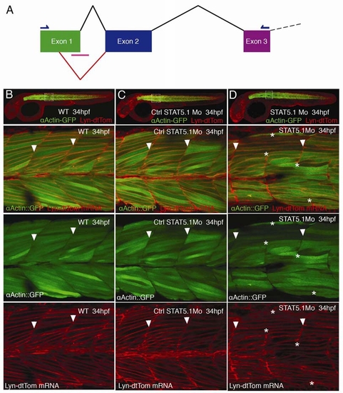
stat5.1 Morphants Show Long Muscle Fibers Extending through Somites |

Unillustrated author statements |
Reprinted from Developmental Cell, 16(2), Liu, Y.H., Jakobsen, J.S., Valentin, G., Amarantos, I., Gilmour, D.T., and Furlong, E.E., A Systematic Analysis of Tinman Function Reveals Eya and JAK-STAT Signaling as Essential Regulators of Muscle Development, 280-291, Copyright (2009) with permission from Elsevier. Full text @ Dev. Cell