
- Title
-
Hedgehog signalling is required for cloacal development in the zebrafish embryo
- Authors
- Parkin, C.A., Allen, C.E., and Ingham, P.W.
- Source
- Full text @ Int. J. Dev. Biol.
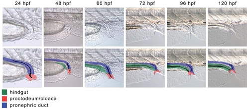
The development of the cloaca in WT embryos. (A-E) Live WT embryos between 24 and 120 hpf. At 24 hpf the proctodeum is visible as a mass of cells at the site of the pronephric opening. The proctodeum surrounds the opening of the pronephric duct at all stages and forms the cloaca. By 48 hpf the posterior gut has reached the cloaca and starts to fuse with it. As the posterior gut becomes canalised from anterior to posterior, the proctodeum invaginates to form the anus. The anal canal, the most distal part of the gastrointestinal tract, is formed by the fusion of the proctodeum with the posterior gut endoderm. By ∼96 hpf the posterior gut lumen is in contact with the ventral edge of the embryo, but is not yet open. At 120 hpf the gastrointestinal tract opens externally, adjacent to the pronephric duct. (G-L) Live WT embryos between 24 and 120 hpf with an overlay showing the identity of various tissues: green, posterior gut; blue, pronephric ducts; red, proctodeum/cloaca. |
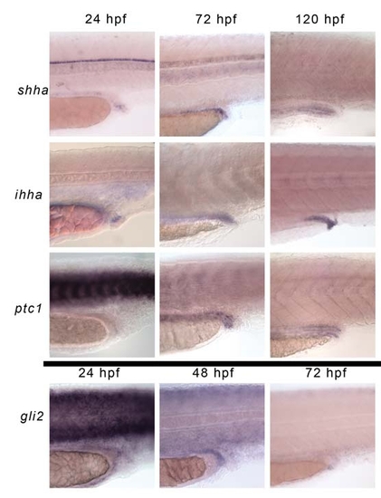
Expression pattern of Hh genes in the developing cloaca. In situ hybridisation stains in WT embryos. (A-C) Transcripts for shha are detected at low levels in the posterior gut endoderm and persist until at least 120 hpf. (D-F) ihha is expressed in the proctodeum from 24 hpf and is expressed in the posterior gastrointestinal tract and proctodeum until at least 120 hpf. (G-H) Before 48 hpf ptc1 transcripts are not up-regulated in the area surrounding the posterior gut, but later ptc1 is expressed at higher levels in the mesodermal layer that surrounds the Hh expressing endoderm. (J-L) At 24 hpf gli2a is expressed in a diffuse pattern around the developing posterior gut, around 48 hpf gli2a expression in the developing posterior gut is at its strongest, with low-level expression on the posterior edge of the pronephric ducts, but not within them. By 72 hpf gli2a is expressed in cells surrounding the anal sphincter, and weakly in a large diffuse area around the urorectal opening. gli2a expression is not detectable in the urorectal region after 96 hpf. |
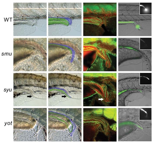
Hh signalling is essential for cloacal development. Bright field images of live Hh pathway mutants and WT siblings (A,B,E,F,I,J,M,N) and confocal scans of embryos stained with α-actin-phalloidin (green) and propidium iodide (red) at 120 hpf (C,G,K,O). Live embryos were injected with fluorescein salt at 72-96 hpf, which accumulates in the gut prior to the opening of the gastrointestinal tract and is then excreted once the gut is functional. Brightfield/fluorescent image merge with the fluorescent channel only shown in the inset (D,H,L,P). The overlay in (B,F,I,N) shows the approximate identity of the relevant tissues, with the posterior gut in green and pronephric ducts in blue. (A,B) WT. By 120 hpf the gastrointestinal tract has fused with the proctodeum and has an independent opening, adjacent to the pronephric ducts. (C) WT. The pronephric ducts have a single narrow opening slightly caudal to the posterior gut. The position where the ducts join is visible in the confocal cross section as an upturned ′U′ shape, with the anterior ducts out of the plane of focus. The posterior gut is surrounded by smooth muscle, and ends with an anal sphincter that is only slightly narrower then the preceding gut. (D) WT embryo excreting fluorescent dye. (E,F) smu. The pronephric ducts are present and are likely to be functional, but the posterior gut is not visible, due to the shape of the embryo and folding of the ventral fins. (G) smu. The pronephric duct caudal to the point where the left and right ducts fuse is much longer in smu mutants because the proctodeum has not opened out. The posterior gut tries to fuse with the proctodeum, but fails, resulting in atresia. (H) smu embryos are completely imperforate (100%, 40/40) and lack peristaltic movement. Application of gentle pressure to the gut moves the dye to the end of the gastrointestinal tract, where it is clear that it is blocked and imperforate. (I,J) syut4 (shha). A channel apparently common between the posterior gut and pronephros (arrow) is suggestive of a fistula between them, but it seems the gut does not fuse correctly with the cloaca leading to imperforate anus. (K) syut4. The posterior gut of syut4 embryos ends blindly dorsal to the proctodeum in most cases. Frequently there is an abortive attempt to fuse with the cloaca leading to the appearance of a fistula between the posterior gut and pronephric duct (arrow). The gastrointestinal tract is completely imperforate – even in instances when there appears to be a fistula. (L) syut4 embryo with imperforate anus/atresia; fluorescent dye can be seen at the blocked end of the gastrointestinal tract in all embryos (100%, 49/49). (M,N) yot (gli2). The pronephric ducts appear to be functional, but are slightly distorted. The gastrointestinal tract is imperforate having failed to fuse with the proctodeum. In this instance peristaltic movements have created a pressure buildup in the distal gastrointestinal tract, causing an outward bulge. (O) yot. The pronephric ducts join together in the correct position, but the usually short single duct appears slightly longer then normal due to the proctodeum failing to open. The posterior gut turns ventrally towards the proctodeum, but fails to fuse with it fully, instead ending blindly adjacent to the pronephric opening. Additionally there is a large reduction in gut girth, probably due to a reduction in smooth muscle in the caudal gut. (P) yot. The majority of yot embryos are imperforate (83%, 15/18), but in the embryos shown here, dye is excreted from the gut through a narrow opening, which is the equivalent of anal stenosis. PHENOTYPE:
|
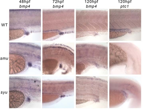
Expression of putative Hh target genes in Hh mutants. Bmp4 and ptc1 expression in Hh pathway mutants and WT siblings. (A-C) WT siblings, 48, 72 and 120 hpf. bmp4 is diffusely expressed in the proctodeum and posterior gut. By 120 hpf expression is restricted to the anus. (D) ptc1 expression in mesenchyme surrounding the posterior gut in a WT sibling. (E-G) smu, 48, 72, 120 hpf. Although the morphological defects in the smu posterior gut alter the shape of the bmp4 expression domain, there are no changes in the levels expressed. (H) There is no ptc1 expression around the cloacal region with the loss of all Hh signal transduction in the smu mutant. (I-K) syu, 48, 72, 120 hpf. Expression of bmp4 in the syu mutant is little different to the WT siblings. (L) Some ptc1 expression surrounding the posterior gut remains after the loss of shha in syu mutants. |

evx1 and prdm1 expression in the cloaca of Hh pathway mutant embryos. (A-D, M-O) WT sibling, 24-120 hpf. At 24 hpf evx1 and prdm1 are expressed in the proctodeum and caudal pronephros. They may also be expressed in the posterior gut at this stage, but it is difficult to determine the presence of endoderm in this area at 24hpf. Expression marked by the arrow may be the proctodeal tissue that fuses with the gastrointestinal tract. Expression in the proctodeum/cloaca is maintained until at least 120hpf. (E-F, P-R) smu, 24-120hpf. Despite morphological changes to the proctodeum and cloaca expression of evx1 and prdm1 are maintained. The altered shape of the expression domain appears to reflect the change in shape of the cloaca seen in live embryos but not a loss of the tissue itself. At 24 hpf–prdm1 expression is not detectable in the putative posterior gut (arrow) whereas evx1 expression is normal. Prdm1 expression in the epidermis persists past 24 hpf in smu mutants (asterix) but not in WT siblings, suggesting that prdm1 is regulated by Hh activity in the epidermis, but not the procotdeum. (I-L, S-U) syu, 24-120hpf. Expression of“evx1 and prdm1 are maintained in the cloaca of syu mutants. At 24hpf the expression of evx1 appears to be weaker than the WT siblings, but at other time points is barely distinguishable from the siblings, whilst prdm1 expression at 24 hpf is normal in syu. Like smu mutants prdm1 expression in the epidermis persists past 24 hpf (asterix). |
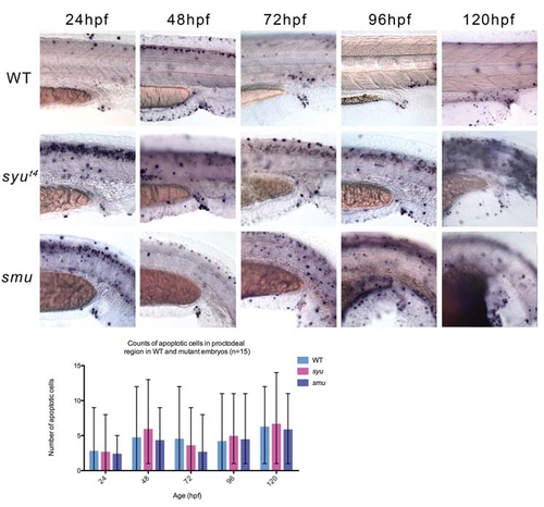
Apoptotic events in the developing posterior gut of wild type and Hh pathway mutants. (A-E) TUNEL staining of WT embryos between 24-120 hpf reveals the normal pattern of apoptosis during the formation of the posterior gut and pronephric ducts. At 120 hpf a number of cells in the cloaca die creating an externally opening posterior gut. (F-J) TUNEL staining of syu mutant between 24-120 hpf. (K-O) TUNEL staining of smu mutants between 24-120 hpf. (P) Counts of apoptotic cells in the mutants and WT siblings reveals that there are large differences in the amount of cell death between embryos of the same age. However, there is no significant difference between the number of apoptotic cells in WT siblings and mutant embryos. |
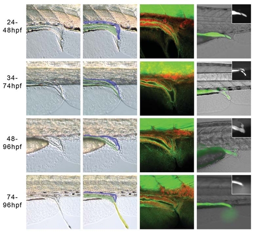
Hh signalling is required between 48-74 hpf for the development of a perforate anus. Embryos treated with 25 μM cyclopmine and imaged live at 120 hpf with bright field illumination, (A,B,E,F, I,J,M,N) or by confocal microscopy following staining with α-actin-phalloidin (green) and propidium iodide (red) (C,G,K,O). The overlay in (B,F,J,N) shows the approximate identity of the relevant tissues, with the posterior gut in green and pronephric ducts in blue. (D,H,L,P) Merged brightfield/fluorecent images of live embryos injected with fluorescein salt at 72-96 hpf, with the fluorescent channel only shown in the inset. (A,B) After treatment from 24-48 hpf, the posterior gut is thinner than in WT embryos, and despite the appearance of a fistula (C), the majority of embryos have a perforate gut, while 33% (4/12) were imperforate and the dye stayed in the blind ended posterior gut (D). (E-H) 34-74 hpf treatment. The posterior gut formed a fistula with the pronephric duct in most embryos, which allowed excretion through a single opening in the proctodeum in around half the embryos (10/18). In the embryos that were not perforate, fluorescein entered the pronephros, but could not be excreted and so moved anteriorly through the pronephric tubes, due to peristaltic pressure (H). (I-L) 48-74hpf treatment. Embryos from this treatment window had a range of phenotypes, 67% (16/24) were perforate, but around half had a fistula to the pronephros (14/24), which itself was not always perforate leading to fluorescein in the pronephric ducts (L). Other embryos had atresia or stenosis. (M-P) 74-96 hpf treatment. The majority of embryos are perforate (90%, 18/20) and the posterior gut musculature affords some control over the excretion of faecal matter. (P) Faecal matter being expelled in a live embryo. |
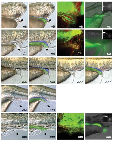
Anal development in mutants with disrupted notochords. Brightfield images of live embryos (A,B,E,F,H,I,L,M) and confocal scans of embryos stained with α-actin-phalloidin (green) and propidium iodide (red) at 120 hpf (C,G,Q). Live embryos were injected with fluorescein salt at 72 hpf,. Brightfield/fluorecent image merge with the fluorescent channel only shown in the inset (D,H,R). The overlay in (B,F,J,L,N,P) shows the approximate identity of the relevant tissues, with the posterior gut in green and pronephric ducts in blue. (A-D) ntl embryos have normal pronephric ducts that open adjacent to the posterior gut. The posterior gut is much thicker in both ntl alleles, but still opens with a normal sized orifice (91% perforate, n11/12). (E-H) flh mutant embryos develop a perforate anus and pronephric duct, which open independently of one another. (87% perforate, n20/23). (I-L) The presence or absence of a notochord near the urogenital opening does not affect its development in bal and doc embryos, which develop a functional anus. (M,N) ctd embryos develop a normal anus and pronephric opening despite undulations in the notochord close to the anus. (O-R) spt embryos have varying pronephric defects, and in some cases lack them altogether; even in the absence of the pronephros (as shown here) the development of the posterior gut is relatively unperturbed (95% perforate, 40/42), although the anal sphincter appears to be absent in most embryos (A,B,E,F,I,J,Q,R) black arrows mark the visible ejection of faecal matter from the anus. PHENOTYPE:
|

Unillustrated author statements PHENOTYPE:
|