
- Title
-
Laser-scanning velocimetry: A confocal microscopy method for quantitative measurement of cardiovascular performance in zebrafish embryos and larvae
- Authors
- Malone, M.H., Sciaky, N., Stalheim, L., Hahn, K.M., Linney, E., and Johnson, G.L.
- Source
- Full text @ BMC Biotechnol.
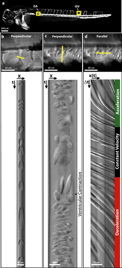
Acquring linescan images from Danio rerio embryos. (a) In silico microangiogram of a 48 hpf wild-type zebrafish embryo. Regions of interest used for acquiring linescan images are boxed (intersomitic vessel = ISV, dorsal aorta = DA). (b and c) A single plane (XY) image (Top) showing the orientation of scan lines used to create perpendicular linescans (Bottom) of the intersomitic vessels (b) and dorsal aorta (c). The boundary between slow moving (elongated) cells and fast moving (compressed) cells reflects a ventricular contraction and is easily observed in the Dorsal Aorta (c). (d) A parallel linescan acquired from the dorsal aorta shows a series of lines whose slope is inversely proportional to cellular velocity. Distinct regions of acceleration, constant velocity, and deceleration are observed. |
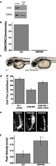
Laser-scanning velocimetry provides functional evidence for the presence of an aortic arch constriction in OSM-deficient embryos. (a) Western blotting for OSM in lysates from control or OSM morpholino-injected embryos harvested at 48 hpf shows nearly complete repression of OSM expression. The abundance of ERK2 is shown as a loading control. (b) Intensity of OSM bands normalized to ERK2 by densitometry for western blots shown in Panel a. (c) OSM morphant embryos at 48 hpf exhibited pericardial edema (arrowhead). (d) Co-injection of OSM mRNA along with OSM morpholino restored cell flux to levels similar to that of control embryos. Cell flux was quantified by scan lines drawn perpendicular to the dorsal aorta above the cloaca. Error bars represent the SEM, n = 3. (e) In silico microangiography of the aortic arch (arrowheads) at 28 hpf shows constricted flow through this region of the outflow tract in OSM-deficient embryos. A region of post-stenotic dilation (arrow), the result of turbulent flow, is occasionally observed. Scale bar is 25 μm. (f) Peak blood cell velocity, measured by laser-scanning velocimetry is increased in OSM-deficient embryos. Error bars represent SEM. Asterisk denotes p = 0.0017 for the two-tailed Student's T-test, n = 3. OSM morpholino #1 was used for the data presented here. Laser-Scanning Velocimetry Data Analyzer smoothing parameters used for trace, velocity, and acceleration smoothing were 5, 10, and 20, respectively. PHENOTYPE:
|

Comparison of cardiac performance measurements obtained at a rostral and caudal position along the dorsal aorta of a 48 hpf embryo. a) In silico microangiogram of a 48 hpf zebrafish embryo showing the location of two scan lines used to acquire velocimetry data from a rostral and caudal location along the dorsal aorta. b-e) Peak velocity (b), peak acceleration (c), stroke volume (d), and cardiac output (e) obtained from rostral and caudal scan lines. Error bars represent the SEM, n=3. |
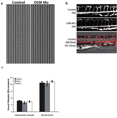
Decreased circulation in OSM-deficient embryos only affects the lumen of the aortic arch Perpendicular line scan images from the intersomitic vessels of control and OSM-morphant embryos clearly illustrate the decreased circulation observed in OSM morphant embryos at 48 hpf (Supplementary Fig. 1a). In silico microangiograms from the tails of 48 hpf control or OSM-deficient embryos show that the dorsal aorta, formed through vasculogenesis, and the intersomitic vessels, which are formed through angiogenesis, are well developed, properly oriented, and make the appropriate connections in both control and OSM morphant embryos (Supplementary Fig. 1b). Because in silico microangiography requires blood cell motion to delineate the path of a vessel, the intensity of the in silico microangiograms from OSM-deficient embryos were less intense than control embryos. To ensure the decreased intensity was not the result of decreased vessel diameters, the diameter of intersomitic vessels and the dorsal aorta were measured by DIC microscopy. Diameters were not different in control embryos of those injected with either of two OSM morpholinos (Supplementary Fig. 1c). PHENOTYPE:
|
|
Unillustrated author statements PHENOTYPE:
|