
- Title
-
laminin alpha 1 gene is essential for normal lens development in zebrafish
- Authors
- Zinkevich, N.S., Bosenko, D.V., Link, B.A., and Semina, E.V.
- Source
- Full text @ BMC Dev. Biol.
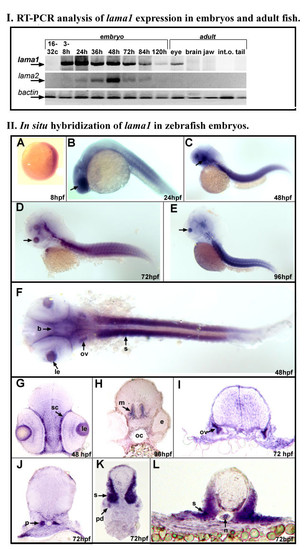
Expression of zebrafish laminin alpha 1 gene. I. RT-PCR analysis of lama1 expression in embryos and adult fish. RT-PCR results for lama1, lama2 and control bactin transcripts are presented as indicated. Embryonic (16-32 cells to 120-hpf) or adult (1 year old) cDNA samples employed in reactions are indicated at the top: lane 1- 16-32 cells, 2- 3-8 hpf, 3- 24 hpf, 4- 36 hpf, 5- 48 hpf, 6- 72 hpf, 7- 84 hpf, 8- 120 hpf embryos; for adult tissues- lane 9 contains products obtained with adult eye cDNA, 10- brain, 11- jaws, 12- internal organs and 13- tail. II. In situ hybridization of antisense lama1 riboprobe in zebrafish embryos. A-F: 8-96 hpf whole zebrafish embryos that were hybridized with lama1 DIG-labeled antisense riboprobe. G-L: Transverse sections of 48-96 hpf zebrafish embryos at the level of the eye (G), brain (H), otic vesicle (I), developing kidney (J), and trunk (K, L). Embryonic stages are indicated at the bottom of the picture. At 8-hpf, expression of the lama1 gene was detected in all embryonic tissues; by 24-hpf, higher levels of transcript were evident in the developing lens (arrows in B-E; le in F and G) and sclera (sc) of the eye, brain (b), somites (s), and otic vesicle (ov), pronephros (p) and pronephric duct (pd), notochord (n). e- eye, m-midbrain. PHENOTYPE:
|
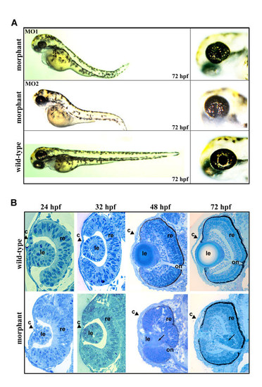
lama 1 knockdown phenotype in zebrafish. A, an overall view of lama1-morphants obtained with MO1- (top) or MO2- oligomers (middle), and control (bottom) embryos at 72-hpf. Enlarged image of a head is provided on the right. Defects in body length, axis curvature and eye structure (irregular pupil and a lack of lens) are easily detectable in lama1 morphants. B, transverse sections at the eye level of control (top row) and lama1-morphant (bottom row) embryos at 24-, 32-, 48- and 72-hpf are presented. An obvious lens degeneration is first notable in 48-hpf morphant eyes. At 72-hpf, small eyes with missing lens and thickened cornea were observed in the morpholino-injected embryos. retina (r), optic nerve (on), lens (le) and cornea (c) are shown. Black arrows in 48- and 72-hpf eyes indicate to the products of lens degeneration. |