
|
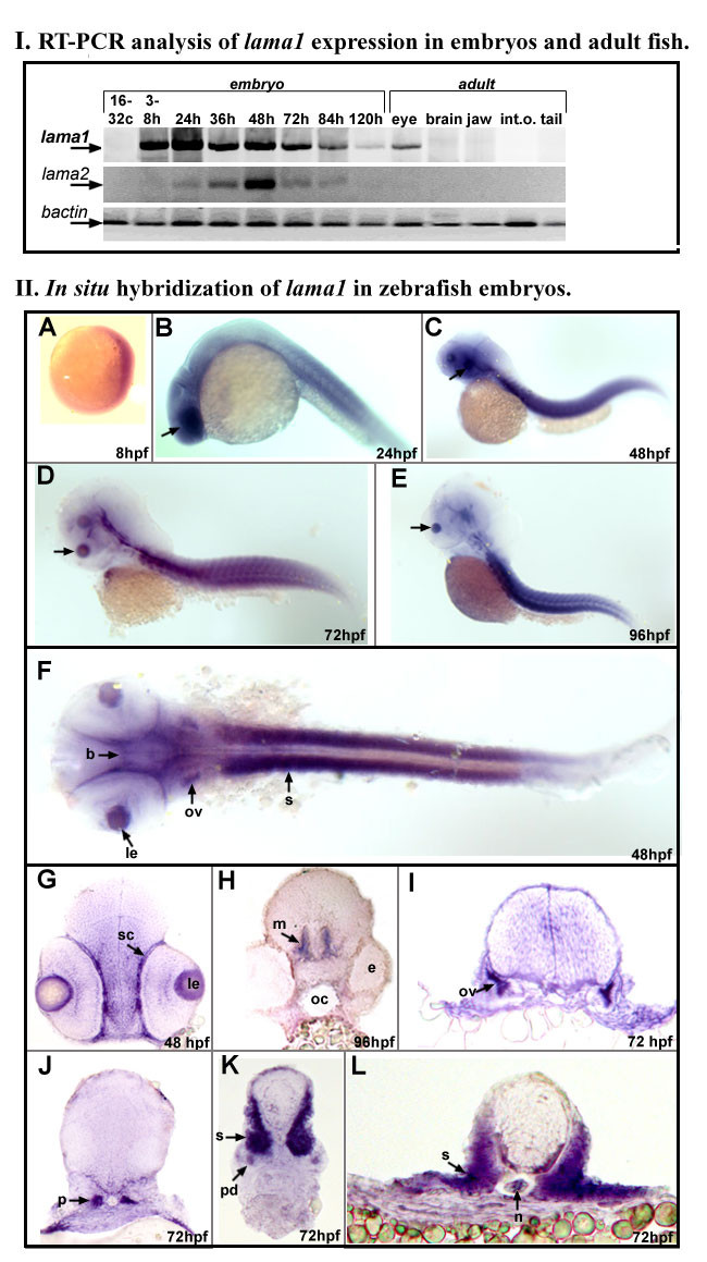
Fig. 3 Expression of zebrafish laminin alpha 1 gene. I. RT-PCR analysis of lama1 expression in embryos and adult fish. RT-PCR results for lama1, lama2 and control bactin transcripts are presented as indicated. Embryonic (16-32 cells to 120-hpf) or adult (1 year old) cDNA samples employed in reactions are indicated at the top: lane 1- 16-32 cells, 2- 3-8 hpf, 3- 24 hpf, 4- 36 hpf, 5- 48 hpf, 6- 72 hpf, 7- 84 hpf, 8- 120 hpf embryos; for adult tissues- lane 9 contains products obtained with adult eye cDNA, 10- brain, 11- jaws, 12- internal organs and 13- tail. II. In situ hybridization of antisense lama1 riboprobe in zebrafish embryos. A-F: 8-96 hpf whole zebrafish embryos that were hybridized with lama1 DIG-labeled antisense riboprobe. G-L: Transverse sections of 48-96 hpf zebrafish embryos at the level of the eye (G), brain (H), otic vesicle (I), developing kidney (J), and trunk (K, L). Embryonic stages are indicated at the bottom of the picture. At 8-hpf, expression of the lama1 gene was detected in all embryonic tissues; by 24-hpf, higher levels of transcript were evident in the developing lens (arrows in B-E; le in F and G) and sclera (sc) of the eye, brain (b), somites (s), and otic vesicle (ov), pronephros (p) and pronephric duct (pd), notochord (n). e- eye, m-midbrain.