
- Title
-
Cre-mediated site-specific recombination in zebrafish embryos
- Authors
- Thummel, R., Burket, C.T., Brewer, J.L., Sarras, M.P. Jr., Li, L., Perry, M., McDermott, J.P., Sauer, B., Hyde, D.R., and Godwin, A.R.
- Source
- Full text @ Dev. Dyn.

Cartoon of HSP70-EGFP-cre and recombination reporter plasmids. A: HSP70-EGFP-cre construct. B: CMV driven hcRFP reporter construct. Gray triangles denote lox511 sites, and the red stop sign denotes the synthetic polyadenylation site and C2 termination site. C: PGK promoter driven lacZ reporter construct. Gray triangles denote loxP sites, and the red stop sign denotes the insertion of the neomycin (or G418) resistance gene and SV40 polyadenylation site. D: CMV-driven EGFP reporter (Cre Stoplight) construct. Gray triangles denote loxH (immediately 3’ to the CMV promoter) and loxP (3’ to the stop sign) sites, and the red stop sign denotes the insertion of the dsRFP gene and bovine growth hormone gene polyadenylation site. B-D: Small bent arrows denote transcriptional start sites, and the EGFP-Cre fusion protein is denoted by the fused green and blue spheres. |

Heat shock regulated expression of EGFP and EGFP-Cre. A: Mosaic transgenic zebrafish (24 hours postfertilization [hpf]) for the HSP70-EGFP construct in the absence of heat shock. B: Mosaic transgenic zebrafish (24 hpf) for the HSP70-EGFP construct heated for 90 sec at 42°C. C: Mosaic transgenic zebrafish (24 hpf) for the HSP70-EGFP-cre construct in the absence of heat shock. D: Mosaic transgenic zebrafish (24 hpf) for the HSP70-EGFP-cre construct heated for 90 sec at 42°C. E: Confocal Z-stack showing a representative HSP70-EGFP-cre Tg/Tg zebrafish at approximately 24 hpf. Brightfield images of the embryos in A-D are shown in the insets. Scale bars = 250 microns in B (applies to inset and A-D), 500 microns in E. |
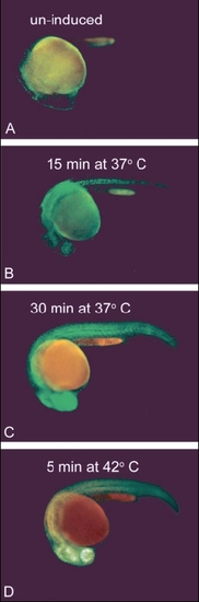
Analysis of heat shock induction schemes for HSP70-EGFP-cre transgenic line. A: Un-induced HSP70-EGFP-cre Tg/+ embryo photographed at 24 hours postfertilization (hpf). B: HSP70-EGFP-cre Tg/+ embryo photographed at 24 hpf after being induced for 15 min at 37°C at 20 hpf. C: HSP70-EGFP-cre Tg/+ embryo photographed at 24 hpf after being induced for 30 min at 37°C at 20 hpf. (D) HSP70-EGFP-cre Tg/+ embryo photographed at 24 hpf after being induced for 5 min at 42°C at 20 hpf. |
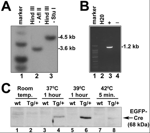
Analysis of HSP70-EGFP-cre transgenic line. A: Southern transfer analysis of HSP70-EGFP-cre transgenic line. B: Reverse transcriptase-polymerase chain reaction analysis of HSP70-EGFP-cre transgenic line. "H20" denotes no DNA control reaction. "+" and "-" denote addition and absence of reverse transcriptase, respectively. C: Western blot detection of EGFP-Cre fusion protein. Lane 1: un-induced wild-type fish; lane 2: un-induced HSP70-EGFP-cre Tg/+ fish; lane 3: wild-type fish heat shock induced for 1 hr at 37°C; lane 4: HSP70-EGFP-cre Tg/+ fish heat shock induced for 1 hr at 37°C; lane 5: wild-type fish heat shock induced for 1 hr at 39°C; lane 6; HSP70-EGFP-cre Tg/+ fish heat shock induced for 1 hr at 39°C; lane 7: wild-type fish heat shock induced for 5 min at 42°C; lane 8: HSP70-EGFP-cre Tg/+ fish heat shock induced for 5 min at 42°C. |

Detection of hcRFP after heat shock induction of EGFP-Cre. A: Lack of detectable expression from the hcRFP reporter construct (depicted at the top of the panel) in mosaic transgenic embryos at 24 hours postfertilization (hpf). B: Detection of expression from final recombination product (depicted at the top of the panel) in mosaic transgenic embryos at 24 hpf. C-F: Detection of hcRFP-positive cells after heat shock induction of EGFP-Cre. C: Brightfield image showing the trunk of an HSP70-EGFP-cre embryo injected with the hcRFP reporter construct. D: EGFP fluorescent image of embryo shown in C, showing remaining EGFP intensity 24 hr post heat shock. E: hcRFP fluorescent image of embryo shown in C. Arrow denotes a cluster of hcRFP-positive cells 24 hr post heat shock. F: Artificial merge of brightfield and fluorescent images. Arrow denotes the cluster of hcRFP-positive cells shown in E. Scale bars = 250 microns in A (applies to A,B and insets). |

Detection of β-galactosidase after heat shock induction of EGFP-Cre. A: Detection of expression from final lacZ recombination product (depicted at the top of the panel) in mosaic transgenic embryos at 24 hours postfertlization (hpf). B: Detection of β-galactosidase-positive (blue) cells after heat induction of a lacZ reporter injected HSP70-EGFP-cre transgenic embryo at approximately 42 hpf. C: Enlargement of boxed region of B. |

Detection of Cre-mediated site-specific deletion by polymerase chain reaction (PCR) analysis. A: Cartoon of two expected unrecombined PCR products. B: Cartoon of expected recombined PCR product. C: Analysis of recombination in EGFP-cre Tg/+ X pCMV-loxH-STOP-loxP-EGFP reporter Tg/+ cross and confirmation of presence of EGFP-cre Tg. "H2O" refers to the water (no DNA) control PCR reaction, and "no hs" refers to genomic DNA isolated from the transgenic cross in the absence of heat shock induction. |