
- Title
-
Histone deacetylase 1 is required for cell cycle exit and differentiation in the zebrafish retina
- Authors
- Stadler, J.A., Shkumatava, A., Norton, W.H., Rau, M.J., Geisler, R., Fischer, S., and Neumann, C.J.
- Source
- Full text @ Dev. Dyn.
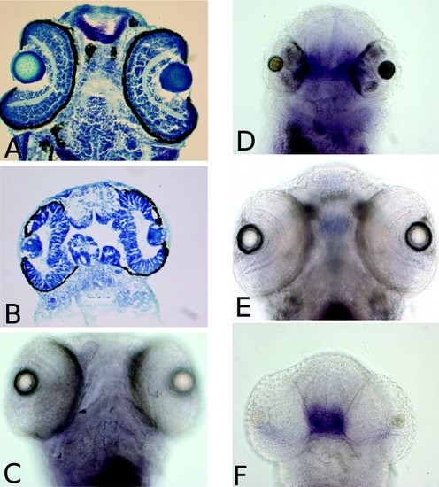
The optic stalk is enlarged at the expense of the retina. A,B: Borreline staining of 96 postfertilization (hpf) wild-type (A) and hdac1 mutant (B), cryosections (dorsal view, anterior up). Note the presence of an overgrown optic stalk in the hdac1 mutant. C-F: Whole-mount in situ hybridization of pax2 (C,D) and vax1 probes (E,F); 120 hpf wild-type (C,E) and hdac1 mutant (D,F) embryos (ventral view, anterior up). EXPRESSION / LABELING:
|
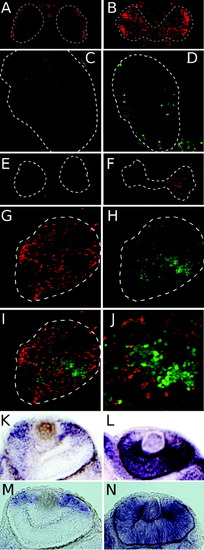
>Loss of hdac1 activity results in failure of retinal cells to exit the cell cycle. A,B: A 2-hr bromodeoxyuridine (BrdU) incorporation in 72 hr postfertilization (hpf) wild-type (A) or hdac1 mutant (B) embryos. C,D: Anti-H3 immunofluorescence on cryosections of 72 hpf wild-type (C) or hdac1 mutant (D) eyes. E,F: Terminal deoxynucleotidyl transferase-mediated deoxyuridinetriphosphate nick end-labeling (TUNEL) staining of apoptotic cells in 72 hpf wild-type (E) and hdac1 mutant (F) embryos. G-J: Wild-type green fluorescent protein (GFP) -expressing cells (green, H,I) were transplanted into hdac1 mutant embryos. BrdU incorporation is shown (red, G,I). J: Higher magnification of the image shown in I. A-J shows immunostaining on cryosections, anterior up. K-N: Whole-mount in situ hybridizations of cyclin D1 RNA (K,L), and cyclin E2 RNA (M,N); 48 hpf wild-type (K,M) and hdac1 mutant (L,N) embryos (anterior right, dorsal up) EXPRESSION / LABELING:
|