
- Title
-
Novel steroidogenic factor-1 homolog (ff1d) is coexpressed with anti-Mullerian hormone (AMH) in zebrafish
- Authors
- von Hofsten, J., Larsson, A., and Olsson, P.E.
- Source
- Full text @ Dev. Dyn.

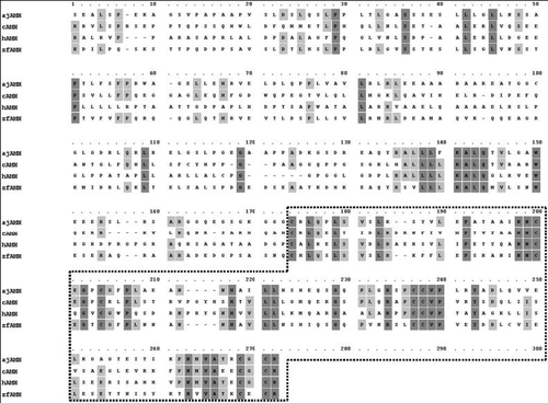
Protein alignment of zebrafish FF1 full-length sequences. The 28 amino acid FTZ-F1 box is underlined. The DNA-binding domain is indicated by a square. The I-box and AF-2 domains are underlined in the ligand binding domain. Consensus amino acids are boxed in dark gray, and amino acids shared by three of four sequences are boxed in light gray. Amino acids identical between ff1b and ff1d are indicated by asterisks. |
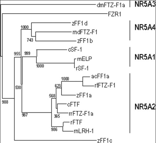
NR5A sequence similarity analysis displayed in a phylogenetic tree. Clades containing subgroups NR5A1, NR5A2, NR5A3, and NR5A4 are indicated. Arctic char FF1a (acFF1a); mouse LRH-1 (mLRH-1); rat SF-1 (rSF-1); mouse ELP (mELP); Rana rugosa FTZ-F1 (rrFTZ-F1); zebrafish FF1b (zFF1b); zebrafish FF1a (zFF1a); zebrafish FF1c (zFF1c); zebrafish FF1d (zFF1d); rat FTF (rFTF); medaka FTZ-F1 (mdFTZ-F1); rainbow trout FTZ-F1 (rtFTZ-F1); chick SF-1 (cSF-1); chick FTF (cFTF); and Drosophila melanogaster ftz-f1 (dmFTZ-F1). The numbers at the base of each clade division represent bootstrap values after 1,000 repeats. |
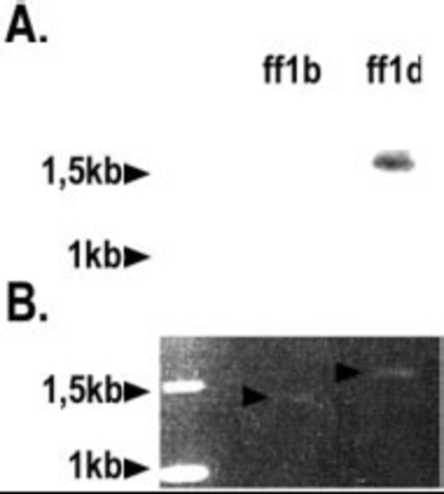
ff1d probe specificity Southern blot. The specificity of the ff1d probe used in subsequent in situ analyses was tested against ff1b and ff1d templates by Southern blot. A: Detection of ff1d template after Southern hybridization. B: Gel loading control. |
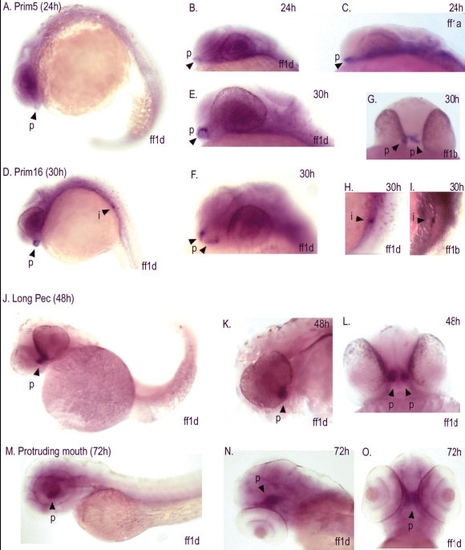
Developmental expression of ff1d detected by whole-mount in situ hybridization. A,B: Detection of ff1d in the pituitary of 24 hr embryos. C: Detection of ff1a in the pituitary of 24 hr embryos. D-F: Detection of ff1d in the pituitary of 30 hr embryos. G: Detection of ff1b in the pituitary of 30 hr embryos. H,I: Detection of ff1d and ff1b in the interrenal of 30 hr embryos. J-L: Detection of ff1d in the pituitary of 48 hr embryos. M-O: Detection of ff1d in the pituitary of 72 hr embryos. p, pituitary; i, interrenal. EXPRESSION / LABELING:
|
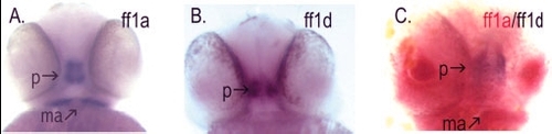
Colocalization of ff1a and ff1d in the pituitary of 48 hr embryos by two-color whole-mount in situ hybridization. A:ff1a expression in pituitary and mandibular arch. B: ff1d expression in pituitary. C: ff1a expression in pituitary and mandibular arch detected with Fast Red, ff1d expression in pituitary detected with nitroblue tetrazolium/5-bromo-4-chloro-3-indolyl phosphate (NBT/BCIP). p, pituitary; ma, mandibular arch. |
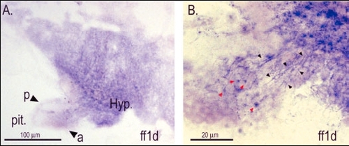
A: ff1d expression in hypothalamus and anterior pituitary at 30 days postfertilization. B: ff1d expression in neurons descending from the hypothalamus to the anterior pituitary. p, posterior pituitary; a, anterior pituitary; black arrowheads, descending neurons; red arrowheads, cells in the anterior pituitary. |
Alignment of anti-Mullerian hormone (AMH) protein from human (hAMH), chick (cAMH), eel (ajAMH), and zebrafish (zfAMH). The transforming growth factor-beta-like region is boxed. Consensus amino acids are boxed in dark gray, and amino acids shared by three of four sequences are boxed in light gray. |

Embryonic expression of ff1a, ff1b, ff1d, and AMH in adult zebrafish, detected by reverse transcriptase-polymerase chain reaction. Stages: O, unfertilized oocytes; 1K, 1K stage; 50% epiboly; 1s, 1 somite; 10s, 10 somites; 28s, 28 somites; P16, Prim 16; LP, Long Pec; 10d, 10 days postfertilization (dpf); 20d, 20 dpf; 30d, 30 dpf. AMH, anti-Mullerian hormone. |

Tissue distribution of ff1a, ff1b, ff1d, and AMH in adult zebrafish, detected by reverse transcriptase-polymerase chain reaction. m, male tissue; f, female tissue. Cont, control; +, plasmid control; -, no template. AMH, anti-Mullerian hormone; 18S, 18 SrRNA. |
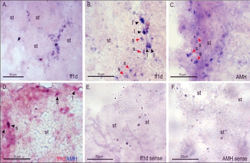
Expression of ff1d and AMH in the testis. A: Overview of ff1d expression in the interstitial region. B: ff1d testicular expression was restricted to interstitial Leydig cells and Sertoli cells. C: AMH expression in the Sertoli cells. D: Double-stained testis tubule showing coexpression of ff1d and AMH. E: Overview of testis section using digoxigenin- (DIG) labeled ff1d sense probe. F: Overview of testis section using DIG-labeled AMH sense probe. st, seminiferous tubule center; l, Leydig cell; s, Sertoli cell; AMH, anti-Mullerian hormone. |
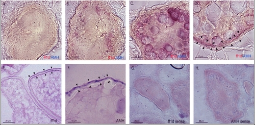
Expression of ff1d and AMH in the ovary. A: Stage Ia follicle lacking AMH and ff1d expression. B: Stage Ib follicle with AMH and ff1d expression in the oocyte. C: Stage II follicle with AMH and ff1d expression in the oocyte. D: Stage III follicle with AMH and ff1d expression in cells follicular layer region. E: Stage III follicle with ff1d expression in cells follicular layer region and the inside of the vitelline envelope. F: Stage III follicle with AMH expression in cells follicular layer region. G: Stage III follicle hybridized with digoxigenin- (DIG) labeled ff1d sense probe. H: Stage III follicle hybridized with DIG-labeled AMH sense probe. Black arrowheads indicate the outer follicular layer region and the inside of the vitelline envelope. AMH, anti-Mullerian hormone. |