
- Title
-
Cloning of zebrafish T-box genes tbx15 and tbx18 and their expression during embryonic development
- Authors
- Begemann, G., Gibert, Y., Meyer, A., and Ingham, P.W.
- Source
- Full text @ Mech. Dev.
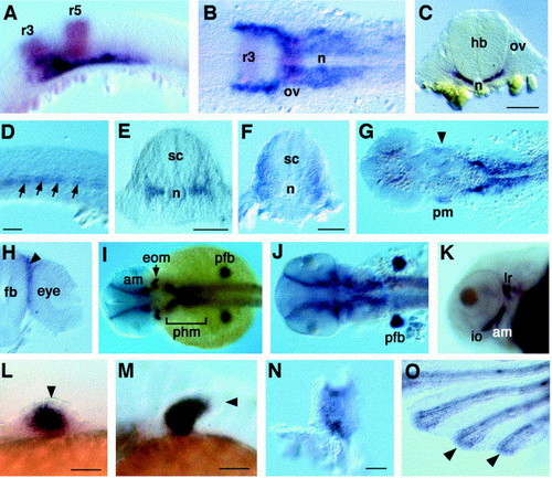
Expression of tbx15 during wild type zebrafish development. (A,B) Double in situ hybridisation showing tbx15 expression (purple) at 17 hpf in the paraxial head mesenchyme, extending rostrally underneath and lateral to rhombomere 3 (r3), counterstained with krox-20 in r3 and r5 (red); (B) dorsal view. (C) At 18 hpf expression is restricted to the paraxial mesenchyme, but excluded from r5 and otic vesicles. (D,E) Somitic expression at 17 hpf in 10–11 rostral somites in the region of the horizontal myoseptum (arrows) expands to the entire somite at 27 hpf (F); (E,F) transverse sections at the level of the fifth somite. (G) Expression is detected in the first pharyngeal arch (at 24 hpf) and (H) in mesenchymal cells separating forebrain and eye. (I,J) tbx15 expression in extraocular and pharyngeal arch muscles, paraxial head mesoderm and pectoral fin bud mesenchyme at (I) 30 hpf and (J) 48 hpf. (K) Strong expression in eye and pharyngeal muscles; ventro-lateral view at 60 hpf. (L–O) tbx15 transcript in developing pectoral fins; expression is exclusively mesodermal, and at 36 h (L) is strong in the medial part of the fin bud and absent from the epidermis (arrowhead); mesodermal expression remains strong throughout the fin bud at (M) 48 hpf and (N) 3 dpf; (O) At 30 dpf tbx15 is strongly expressed in the margins and distal tips of lepidotrichs (arrowheads). (A,D,K–N) lateral views, (B,G,I,J) dorsal views, (C,E,F,H) transverse sections, (O) ventral view. am, adductor mandibulae; eom, extraocular muscles; fb, forebrain; hb, hindbrain; io, inferior oblique; lr, lateral rectus; n, notochord; ov, otic vesicle; pfb, pectoral fin bud; phm, paraxial head mesenchyme; pm, pharyngeal arch mesenchyme; r, rhombomere; sc, spinal cord; Scale bars, 100 μm. EXPRESSION / LABELING:
|

Expression of tbx18 during wild type zebrafish development. (A–C) Somitic expression of tbx18. (A) At 19 hpf expression is strong in the anterior halves of posterior somites (arrowhead); more mature somites express tbx18 above and below the horizontal myoseptum (arrow). (B) Higher magnification, 24 hpf; expression is restricted to the median part of each somite; a transverse section (C) at the level indicated by the arrow reveals the restriction of expression to the somites. (D,E) The ventral neuroectoderm expresses tbx18 at 24 hpf in the medial longitudinal fascicles; r3 is indicated by krox-20 expression. (F,G) tbx18 transcript is detected at 30 hpf in the paraxial head mesenchyme, flanking the otic vesicles medially and dorsally. Note expression in the pectoral fin mesenchyme. (H–J) tbx18 transcript in the developing heart at 36 hpf (H), 48 hpf (I) and 4 dpf (J); strong expression is observed in the septum transversum (arrow); arrowheads point to sites of tbx18 expression in atrium and sinus venosus. (K,K′) Ventral views of the head at 40 hpf; tbx18 is expressed anterior to and surrounding the mouth (bracket indicates region depicted magnified in a different embryo in K′) and in an area flanking the olfactory epithelium; (L) Ventral view of the head of a 3-day-old embryo (lower jaw removed), showing tbx18 expression in the palate (arrow) and in paraxial mesoderm between notochord and ear. (M–Q) tbx18 expression in developing pectoral fins; expression is initiated shortly before 30 hpf (M) in the median part of the fin bud mesenchyme and expands to the entire mesenchyme by 48 hpf (N); (O) at 3 dpf expression is stronger in the anterior mesenchyme and the mesenchymal margin; at 4 dpf (P) expression is restricted to cells bordering the non-expressing apical fold; (Q) pectoral fin at 30 dpf; expression of tbx18 in the interradial zone (asterisks); dark spots on the pectoral fin rays are melanocytes. Distal is to the left, anterior to the top.(C,E,G) transverse sections, (A,B,D,H,I,M–P) lateral views, (J–L,Q) ventral views, (F) dorsal view. a, atrium; hb, hindbrain; m, mouth; mb, midbrain; mlf, medial longitudinal fascicle; n, notochord; oe, olfactory epithelium; ov, otic vesicle; pfb, pectoral fin bud; r, rhombomere; sep, septum transversum; sc, spinal cord; sv, sinus venosus; Scale bars, 100 μm. EXPRESSION / LABELING:
|

Unillustrated author statements EXPRESSION / LABELING:
|
Reprinted from Mechanisms of Development, 114(1-2), Begemann, G., Gibert, Y., Meyer, A., and Ingham, P.W., Cloning of zebrafish T-box genes tbx15 and tbx18 and their expression during embryonic development, 137, Copyright (2002) with permission from Elsevier. Full text @ Mech. Dev.