
|
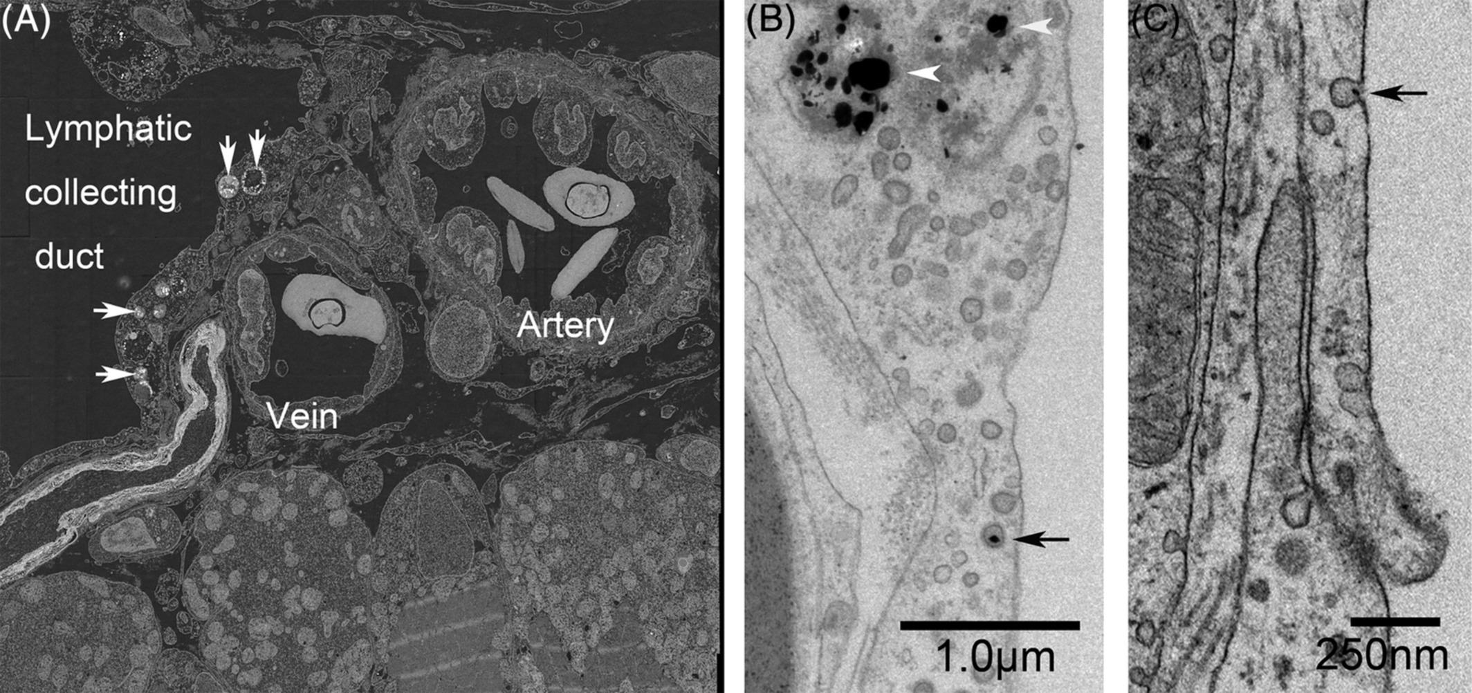
Fig. 5 Lateral lymphatic duct labeled with cinnabar (HgS) and collaterally located subcutaneous artery and vein in the trunk of adult zebrafish. A, Back‐scatter image from the reflection transmission electron microscope revealed that electron‐dense particles (cinnabar; white arrows) accumulated in the endothelia lining the lateral lymphatic duct but not in those of the arteries or veins. B and C, In higher‐magnification views of the lymphatic endothelium, accumulation of dense particles can be seen in the lysosomes (white arrowheads), and a single particle (black arrow) is associated with a small pit (or caveola) and a coated pit