
- Title
-
The role of tolloid/mini fin in dorsoventral pattern formation of the zebrafish embryo
- Authors
- Connors, S.A., Trout, J., Ekker, M., and Mullins, M.C.
- Source
- Full text @ Development
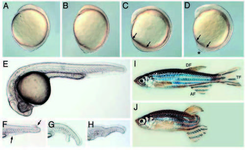
Morphological defects visible in live mini fin mutant embryos. (A,B) 1-somite-stage mfn mutant (B) displays an oblong shape relative to wild type (A). 5- somite-stage wild-type (C) and mfn (D) mutant embryos. Arrows denote the tail bud length and the asterisk marks a protrusion of the tail bud seen in the mutant (D). 24 hpf wild type (E) and tails of mfn mutants exhibiting (F) a partial loss of tail fin (between the arrows) that extends to the dorsal side, (G) partial loss of the ventral fin with a bifurcation in the tail, (H) near complete absence of ventral fin and a kink in the tail. This occasionally observed kink is associated with a disruption in the notochord, which is also observed in the more strongly dorsalized mutant lost-a-fin (Solnica-Krezel et al., 1996, our unpublished observations). (I,J) mfn homozygous adult fish. Homozygous adult exhibiting a near wild-type appearance (I). Note that a pigmented stripe in the tail is disrupted in this mutant, a trait characteristic of mfn homozygous fish. Other adult mutants may display a partial or full (J) loss of their tail fin. All are lateral views. (A-D) dorsal to the right, (E-J) dorsal to the top. AF, anal fin; TF, tail fin; DF, dorsal fin. PHENOTYPE:
|
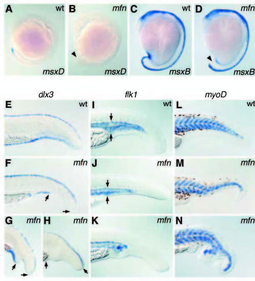
Absence or reduction of three ventral tail cell types in mfn mutants. msxD expression in 8-somite-stage wild-type (A) and mfn mutant (B) embryos. msxB expression in 14-somite-stage wild-type (C) and mfn mutant (D) embryos. msxB is also expressed in other more anterior cell types. dlx3 expression in 24 hpf wildtype (E) and mutant (F-H) embryos. flk1 expression in 24 hpf wild-type (I) and mutant (J,K) embryos. Note reduced flk1 in mfn (arrows). myoD expression in 32 hpf wild-type (L) and mutant (M,N) embryos. Arrowheads in B and D denote reduced/absent gene expression in the mutant. (F-H) Ventral dlx3 expression is absent between the arrows. All are lateral views. (A-D) Dorsal is to the right, anterior to the top. (E-N) Dorsal is to the top, anterior to the left. EXPRESSION / LABELING:
|
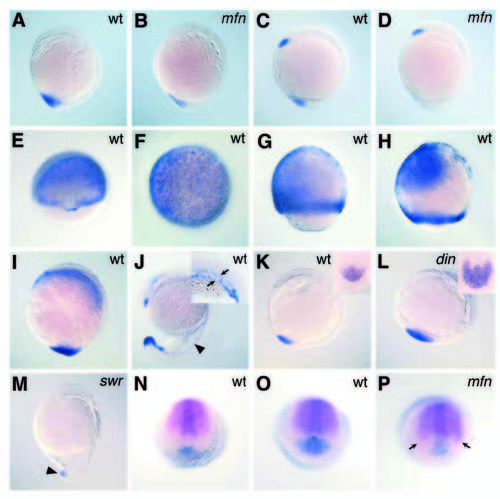
tld expression pattern and altered tld, chd and bmp4 expression in mutant embryos. eve1 expression in bud stage wild-type (A) and mfn mutant (B) embryos. bmp4 expression in 1-somite-stage wild-type (C) and mfn mutant (D) embryos. tld expression in wild-type embryos at 55% epiboly (E), 70% epiboly (F,G), 85% epiboly (H), bud-stage (I) and 20-somite-stage (J) wild-type embryos. Arrowhead in J points to tld expression in the presumptive vasculature. Inset in J is a higher magnification showing the posterior trunk of a slightly older embryo with tld expression marking the prospective artery and vein (arrows). tld expression at 5 somites in wild-type (K), chordino (L) and swirl (M) mutant embryos. Insets in K-L are higher magnification caudal views of tld expression, anterior to the top. Reduced tld expression (arrowhead) in a swirl mutant. Double in situ of chd (magenta) and bmp4 (blue) expression in a bud-stage wild-type embryo (N). Double in situ of chd (magenta) and tld (blue) expression in bud-stage wild-type (O) and mfn mutant (P) embryos. (P) Expanded chd expression (arrows) in a mfn mutant. (A-D, G-M) Lateral views, dorsal to the right. (E) Dorsal view, slightly tilted downward. (F) Animal pole view. (N-P) Vegetal pole view, anterior/dorsal to the top. EXPRESSION / LABELING:
|