
- Title
-
Mutations affecting craniofacial development in zebrafish
- Authors
- Neuhauss, S.C., Solnica-Krezel, L., Schier, A.F., Zwartkruis, F., Stemple, D.L., Malicki, J., Abdelilah, S., Stainier, D.Y., and Driever, W.
- Source
- Full text @ Development
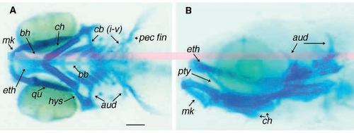
Pharyngeal skeleton of a day- 5 zebrafish larva stained for cartilage with Alcian blue. (A) Ventral view. Individual cartilaginous elements of the pharyngeal skeleton are identifiable. The two trabeculae fuse medially to form the ethmoid plate (eth). First arch derivatives are Meckel’s cartilage (mk) and quadrate (qu). Second arch derivatives are basihyal (bh), ceratohyal (ch), hyosymplectic (hys). Gill arch derivatives are basibranchials (bb) and ceratobranchials I-V (cb i-v). Also labeled are cartilages of the pectoral fins (pec fin). (B) Lateral view reveals the pterygoid process of the quadrate (pty) and the auditory capsule (aud). Scale bar is 100 μm. |

Mutations affecting the layout of the pharyngeal skeleton. (A-H) Lateral view of a living day-5 larvae. (I-P) Ventral view of Alcian blue stained larvae. (A,I) Wild-type embryos. (B,J) little Richardm433. (C,K) mother superiorm188. (D,L) thinnerm214. (E,M) quadrom271. (F,N) nocyranom579. (G,O) bielakm429. (H,P) mont blancm610. For details, see text. wt, wild-type; bh, basihyal; cb, ceratobranchial; ch, ceratohyal; and eth, ethmoid plate. Scale bar, 100 μm. PHENOTYPE:
|
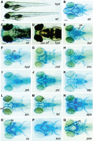
Mutations affecting chondrogenesis. (A) Mutants are shorter and have reduced jaw structures. Wild-type (bottom) and mutant mr hyde205 larva (top) at day 5 of development. The mutant is shorter and has a reduced jaw. Wild-type embryos have an inflated swim bladder. (C) Dorsal view of wild-type day-5 larva. (D) Dorsal view of a day-5 mr hyde205 larva. Notice the rostral position of the eyes and the smaller and bent pectoral fins. (B,E-Q). Ventral views of Alcian blue stained larvae at day 5 of development. (B) Wild-type larva. (E-M) Mutant larvae defective in cartilage formation. Skeletal elements appear smaller and are weakly stained. (E) bulldog494, the ceratobranchials are present but smaller; (F) mr hydem205; (G) roundm713; (H) crusherm299; (I) jekyllm151; (J) zhivagom315; (K) stumpfm365; (L) strangelovem617 and (M) feelgoodm662. N-Q depict mutant larvae that display stunted growth of skeletal elements. (N) apparatchikm364; (O) rieuxm526 (notice the thicker and shorter ceratohyal) and (P) kimblem533. (Q) In postdocm485 the pharyngeal skeleton is reduced and the ceratohyal is bent ventrally. The pectoral fin is reduced. wt, wild-type; ch, ceratohyal; cb, ceratobranchials; mk, Meckel’s cartilage; pec fin; pectoral fin and qu, quadrate. Scale bars, 100 μm. PHENOTYPE:
|
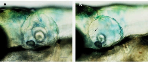
Lateral view of the ear at day 6 of development. (A) In the wildtype the semicircular canals can be seen. (B) Ear of a jekyllm151 larva. The epithelial projections are formed but do not elongate and fail to form the semicircular canals. Arrow points to the cranial projection. Scale bar 50 μm. PHENOTYPE:
|
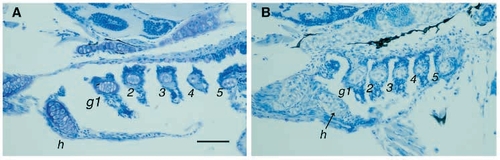
Sagittal sections of the gill arch region of day-5 larvae. Sections were stained with methylene blue. (A) Wildtype. (B) jekyllm151. Notice that the whole gill region is more compressed and that there is a lack of purple staining surrounding the cartilage cells. h, hyoid arch; g1-5, gill arches. Scale bar, 50 μm. PHENOTYPE:
|

Mutations affecting the spatial relationship between elements of the pharyngeal skeleton. Lateral view of live day-5 larvae (A,C,E,G) and their corresponding Alcian blue staining (B,D,F,H). (A,B) Wild type (wt). (C,D) haim114. (E,F) grossmaulm643. (G,H) spitzmaulm636. For details see text. Scale bar, 100 μm. PHENOTYPE:
|

Ventral view of Alcian blue stained day-5 mutant embryos, that have not been assigned to any of the three groups. (A) Wildtype. (B) captain hookm169. (C) maggotm350. (D) white Breadm384. (E) brakm452. (F) An example of a mutant that was initially characterized as having a non-specific delay in development. Alcian blue staining reveals that the ceratohyal is malformed and does not reach the midline. ch, ceratohyal; eth, ethmoid plate; mk, Meckel’s cartilage and qu, quadrate. Scale bar, 100 μm. |
