
- Title
-
Inner Ear and Muscle Developmental Defects in Smpx-Deficient Zebrafish Embryos
- Authors
- Ghilardi, A., Diana, A., Bacchetta, R., Santo, N., Ascagni, M., Prosperi, L., Del Giacco, L.
- Source
- Full text @ Int. J. Mol. Sci.
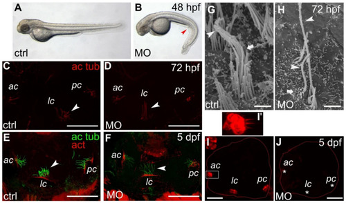
Phenotypical and ear defects in smpx zebrafish morphants (MO). (A,B) representative phenotype caused by the lack of Smpx. Control-MO (A) and smpx-MO (B) injected embryos. The red arrow points to the downward trunk/tail curvature indicative of cilia disfunction. (C–F) kinocilia of the anterior (ac), lateral (lc) and posterior (pc) cristae of the inner ear of 72 hpf (C,D) and 5 dpf (E,F) larvae injected with ctrl- (C,E) and smpx-MO (D,F). Cristae kinocilia are stained with the antibody against acetylated tubulin (ac tub); phalloidin is used to label the actin (act) of the stereocilia. White arrowheads indicate the different ‘posture’ of the lateral crista kinocilia. (G,H) SEM images of the ciliary bundle in the control larvae (G) and smpx morphants (H). The arrowhead indicates the kinocilium, with the arrow the stereocilia. (I,J) representative confocal images of the otic cavity injected with FM4-64, labeling the inner ear hair cells in the control larvae ((I), n = 4) and smpx morphants ((J), n = 7). The asterisks indicate the lack of signal in the ac, lc and pc of the Smpx-deficient larvae, suggestive of an impaired mechanotransduction. (I’) magnified view of the anterior crista in (I) (white rectangle). Scale bars = 50 μm in (C–F); 25 μm in (I,J); 1 μm in (G,H). EXPRESSION / LABELING:
|
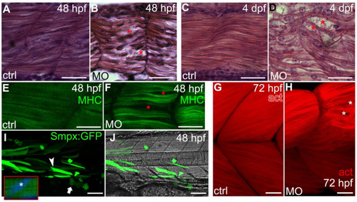
smpx knockdown disturbs the proper fiber arrangement during development. (A–D) side views, anterior left, dorsal top. Paraffin sections of Control-MO (A,C) and smpx-MO (B,D) injected embryos at 48 hpf (A,B) and 4 dpf (C,D). The red asterisks indicate alterations in the muscle array. (E–H) side views, anterior left, dorsal top. Confocal Z-stacks taken from whole-mount embryos (48 hpf) and larvae (72 hpf) labelled with (E,F) myosin heavy chain (MHC) antibody for slow fibers and (G,H) phalloidin (act) for actin filaments of the fast fibers. The red (F) and white (H) asterisks indicate alterations in the normal muscle array (controls in (E,G), respectively). (I,J) representative lateral view of an embryo injected with the construct encoding the Smpx:GFP chimera (see Figure S1A for construct details); muscle fibers (arrowhead) and epithelial cells (arrow) are both uniformly painted with no accumulation of Smpx:GFP in the nuclei. Inset: magnification of a slow fiber with the nucleus (asterisk) labelled with DAPI (blue). Anterior left, dorsal top. Scale bars = 25 μm in (A–D); 20 μm in (E,F); 20 μm in (G,H); 100 μm in (I,J). |