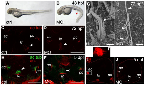
Phenotypical and ear defects in smpx zebrafish morphants (MO). (A,B) representative phenotype caused by the lack of Smpx. Control-MO (A) and smpx-MO (B) injected embryos. The red arrow points to the downward trunk/tail curvature indicative of cilia disfunction. (C–F) kinocilia of the anterior (ac), lateral (lc) and posterior (pc) cristae of the inner ear of 72 hpf (C,D) and 5 dpf (E,F) larvae injected with ctrl- (C,E) and smpx-MO (D,F). Cristae kinocilia are stained with the antibody against acetylated tubulin (ac tub); phalloidin is used to label the actin (act) of the stereocilia. White arrowheads indicate the different ‘posture’ of the lateral crista kinocilia. (G,H) SEM images of the ciliary bundle in the control larvae (G) and smpx morphants (H). The arrowhead indicates the kinocilium, with the arrow the stereocilia. (I,J) representative confocal images of the otic cavity injected with FM4-64, labeling the inner ear hair cells in the control larvae ((I), n = 4) and smpx morphants ((J), n = 7). The asterisks indicate the lack of signal in the ac, lc and pc of the Smpx-deficient larvae, suggestive of an impaired mechanotransduction. (I’) magnified view of the anterior crista in (I) (white rectangle). Scale bars = 50 μm in (C–F); 25 μm in (I,J); 1 μm in (G,H).
|
