FIGURE SUMMARY
Title

Biomechanical signaling within the developing zebrafish heart attunes endocardial growth to myocardial chamber dimensions

Authors
Bornhorst, D., Xia, P., Nakajima, H., Dingare, C., Herzog, W., Lecaudey, V., Mochizuki, N., Heisenberg, C.P., Yelon, D., Abdelilah-Seyfried, S.
Source
Full text @ Nat. Commun.

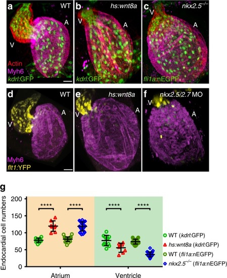
Overexpression of Wnt8a or loss of Nkx2.5 causes a shift in endocardial chamber proportions. ac Reconstruction of confocal z-stacks show the endothelial-specific transgenic reporter lines Tg(kdrl:EGFP)s843 or Tg(fli1a:nEGFP)y7 (green), Phalloidin 568-stained Actin (red) and anti-Myh6 labeling of the myocardial atrial chamber (magenta). Compared to (a) WT, (b) overexpression of Wnt8a, or (c) the nkx2.5vu179 mutation, causes a relative shift of endocardial chamber dimensions. df Reconstructions of confocal z-stacks shows that the arterial endothelial transgene Tg(flt1:YFP)hu4624 marks the ventricular chamber whereas the myocardial atrial marker anti-Myh6 labels the atrial chamber. Compared to (d) WT, (e) overexpression of Wnt8a, or (f) loss of Nkx2.5/Nkx2.7 results in relative shifts of chamber dimensions as indicated by the ventricular-specific expression of Tg(flt1:YFP)hu4624 within the endocardium. A, atrium; V, ventricle. Scale bars, 30 μm. g Quantifications of endocardial cell numbers in WT (kdrl:GFP n = 11 hearts, fli1a:nEGFP n = 27 hearts), Tg(hsp70l:wnt8a-GFP)w34 (n = 8 hearts), or nkx2.5vu179 mutants (n = 27 hearts) reveal that atrial endocardial cell numbers are significantly increased and ventricular endocardial cell numbers are significantly reduced compared to WT. Mean values ± SD are shown. Two-way ANOVA was used to compare each condition with its WT control in each individual chamber (****p < 0.0001)

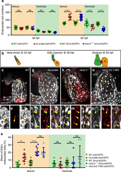
Atrial endocardial cell proliferation is increased upon Wnt8a overexpression or in nkx2.5vu179 mutants. a Quantifications of endocardial cell numbers at 30 hpf reveals that there is no increase of atrial endocardial cells in Tg(hsp70l:wnt8a-GFP)w34 (n = 9 hearts), or nkx2.5vu179 mutant embryos (n = 10 hearts) compared with WT (kdrl:GFP n = 10 hearts, fli1a:nEGFP n = 9 hearts) at this stage. At the later heart ballooning stage (52 hpf), a significant increase of endocardial atrial cell numbers has occurred upon Wnt8a overexpression [Tg(hsp70l:wnt8a-GFP)w34 (n = 8 hearts)] or in nkx2.5vu179 mutants (n = 27 hearts) compared to WT (kdrl:GFP n = 11 hearts, fli1a:nEGFP n = 27 hearts). Mean values ± SD are shown. Two-way ANOVA was used to compare each condition with its WT control in each individual chamber (ns: not significant,****p < 0.0001). b Schematic model of the EdU assay used. WT and Tg(hsp70l:wnt8a-GFP)w34 transgenic embryos were heat shocked at 24 hpf. Embryos of all genotypes were injected with EdU into the circulatory system at 30 hpf and analyzed at 52 hpf. c, g, k, o Reconstructions of confocal z-stacks showing representative hearts at 52 hpf of (c) WT, (g) upon Wnt8a overexpression, (k) in nkx2.5vu179 mutants, or (o) in nkx2.5/nkx2.7 double morphants. Endocardial tissue is marked by Tg(kdrl:EGFP)s843 or Tg(fli1a:nEGFP)y7 reporters (white) and proliferative cells are marked by EdU incorporation (red). A, atrium; V, ventricle. Scale bars, 30 μm. df, hj, ln, pr Shown are magnifications of insets (yellow boxes) with kdrl:GFP or fli1a:nEGFP/EdU double positive cells (yellow arrowhead). Single channel with kdrl:GFP or fli1a:nEGFP (d, h, l, p), EdU incorporation (e, i, m, q) and the merge of both channels (f, j, n, r). Scale bars, 10 μm. s Quantifications of the share of EdU + atrial or ventricular endocardial cells relative to the total number of endocardial cells within the respective cardiac chamber in WT (kdrl:GFP n = 8 hearts, fli1a:nEGFP n = 13 hearts), upon Wnt8a overexpression (n = 9 hearts), nkx2.5vu179 mutants (n = 8 hearts), or nkx2.5/nkx2.7 double morphants (n = 8 hearts). Upon Wnt8a overexpression or loss of Nkx2.5 or Nkx2.5/Nkx2.7, proliferation significantly increases in the developing atrial endocardium. Mean values ± SD are shown. Two-way ANOVA was used to compare each condition with its WT control in each individual chamber (ns: not significant; ***p < 0.001, ****p < 0.0001)

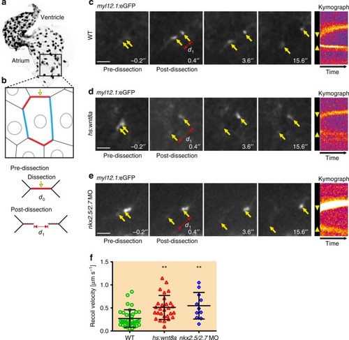
Endocardial tissue tension increases upon Wnt8a overexpression or loss of Nkx2.5/Nkx2.7. a, b Schematic model illustrating the endocardial region in which the laser cuts were performed. Local laser cut (yellow arrow) of the subcortical actomyosin network within the shorter membrane compartment (red) which is oriented perpendicular to the direction of intra-cardiac blood flow at time point d0 causes an actomyosin recoil (d1). ce Time lapse sequences following a laser cut within the endocardium of 40 hpf embryos with the Tg(act2:myl12.1-EGFP)e2212 transgenic reporter that marks the actomyosin network. Yellow arrows show the actomyosin recoil distance within 0.4 ms upon laser dissection. Kymographs indicate the temporal recoil of the actomyosin network along the membrane compartment where the laser cut was performed. The recoil of the open junction ends is visualized with the fluorescence intensity and the initial opening of the junction is marked by yellow arrowheads. d, e Time lapse analyses demonstrate a larger opening of the junction (d1) and a faster recoil of the actomyosin network (yellow arrowheads in the kymograph) upon Wnt8a overexpression or loss of Nkx2.5/Nkx2.7 following laser cuts. Scale bars, 20 μm. f Comparison of initial recoil velocities (μm per sec) which are lower in WT (n = 37 hearts) compared with Wnt8a overexpressing embryos (n = 28 hearts) or upon loss of Nkx2.5/Nkx2.7 (n = 11 hearts). Mean values ± SD are shown. One-way ANOVA was used to compare each condition with WT (**p < 0.001)

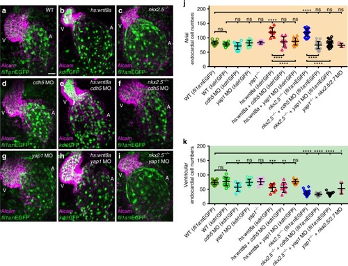
Loss of Cadherin-5 or Yap1 prevents endocardial cell number increases during atrial chamber expansion. a–i Reconstructions of confocal z-stacks of zebrafish hearts at 52 hpf expressing the endocardial reporter Tg(kdrl:EGFP)s843 or Tg(fli1a:nEGFP)y7 (green) and immunolabeling against the myocardial marker Alcam (magenta). j Quantifications of endocardial cell numbers at 52 hpf. b, j Upon Wnt8a overexpression (n = 7 hearts), atrial endocardial cell numbers increase. c, j Similarly, in nkx2.5vu179 mutants (n = 27 hearts), endocardial cell numbers increase within the atrium. d, j MO-mediated knock down of Cadherin-5 (Cdh5) (n = 7 hearts) does not cause a reduction of endocardial cell numbers within the atrium. e, j Loss of Cdh5 prevents increased atrial endocardial cell numbers upon overexpression of Wnt8a (n = 8 hearts). f, j Loss of Cdh5 suppresses increased atrial endocardial cell numbers in nkx2.5vu179 mutants (n = 19 hearts). g, j Loss of Yap1 via MO-mediated knock down (n = 14 hearts) or in yap1fu48 mutants (j, k) does not affect ventricular or atrial endocardial cell numbers. h, j Knock down of Yap1 in Wnt8a overexpressing embryos normalizes ventricular and atrial endocardial cell numbers (n = 8 hearts). i, j Loss of Yap1 rescues atrial endocardial cell numbers in nkx2.5vu179 mutants (n = 17 hearts). j A loss of Nkx2.5/Nkx2.7 in yap1fu48 mutants also rescues atrial endocardial cell numbers. A atrium, V ventricle. Scale bars, 30 μm. j, k Quantifications of endocardial cell numbers in atrium and ventricle. Mean values ± SD are shown. Two-way ANOVA was used to compare each condition with its WT control in each individual chamber (ns not significant; **p < 0.01;***p < 0.001; ****p < 0.0001)

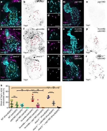
Yap1 nuclear localization within the atrial endocardium increases upon chamber expansion. a, e, i, m, o, q Reconstructions of confocal z-stacks of zebrafish hearts at 52 hpf expressing the endocardial reporters Tg(kdrl:EGFP)s843 or Tg(fli1a:nEGFP)y7 (cyan) and immunolabeling against zebrafish Yap1 (magenta). b, f, j, n, p, r Yap1 immunolabeling is inverted in black/white and endocardial cells which show co-localization with Yap1 labeling are marked with red asterisks. A atrium, V ventricle. Scale bars, 30 μm. c, d, g, h, k, l Magnifications of single confocal XY section planes (yellow box in a, e, i) are shown in c, g, k and, in d, h, l, only Yap1 immunolabeling is shown. Endocardial cells, labeled by Tg(kdrl:EGFP)s843 or Tg(fli1a:nEGFP)y7, that show co-localization with Yap1 are indicated with an asterisk. Scale bars, 30 μm. s Quantifications of the share of Yap1-positive endocardial cells relative to the total number of endocardial cells within the atrium. Upon Wnt8a overexpression (n = 6 hearts) or loss of Nkx2.5 (n = 3 hearts), the share of Yap1-positive endocardial cell numbers significantly increases within the developing atrial endocardium. Loss of Cadherin-5 (Cdh5) changes the share of Yap1-positive endocardial cells among Wnt8a overexpressing (n = 9 hearts) or nkx2.5vu179 mutant embryos (n = 10 hearts) to WT levels. Knock down of Yap1 in all conditions leads to a massive reduction of Yap1-positive endocardial cells within the atrium (yap1 MO: n = 11 hearts; yap1 MO + hs:Wnt8a: n = 9 hearts; yap1 MO + nkx2.5vu179 mutant: n = 11 hearts). Mean values ± SD are shown. One-way ANOVA was used to compare each condition with its WT control (ns not significant, **p < 0.01;***p < 0.001; ****p < 0.0001)

Acknowledgments
This image is the copyrighted work of the attributed author or publisher, and ZFIN has permission only to display this image to its users. Additional permissions should be obtained from the applicable author or publisher of the image. Full text @ Nat. Commun.