FIGURE SUMMARY
Title

H3K27me3-mediated silencing of structural genes is required for zebrafish heart regeneration

Authors
Ben-Yair, R., Butty, V.L., Busby, M., Qiu, Y., Levine, S.S., Goren, A., Boyer, L.A., Burns, C.G., Burns, C.E.
Source
Full text @ Development


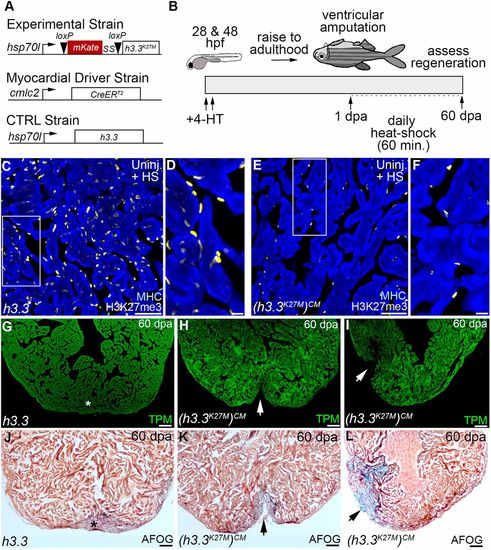
  • Myocardial H3K27 tri-methylation is required for zebrafish heart regeneration in vivo. (A) Schematics depicting driver and inducible transgenes used for Cre/loxP-mediated myocardial-specific expression of Tg(hsp70l:h3.3) (wild-type control) or Tg(hsp70l:h3.3K27M)CM (mutant) during the regenerative window. (B) Schematic depicting the experimental strategy for heart regeneration analyses. Double-transgenic embryos were exposed to 4-HT at 24 h post-fertilization (hpf) and 48 hpf to recombine the mutant transgene specifically in the myocardium. Zebrafish were raised to adulthood and their ventricles resected followed by a 24 h recovery period and daily 1 h heat-shocking for 60 days prior to analysis. (C-F) Sections of uninjured zebrafish ventricles co-immunostained for the cardiomyocyte marker, myosin heavy chain (blue) and H3K27me3 (yellow). Boxed regions in C and E are shown in D and F. Myocardial and non-myocardial cells displayed strong H3K27me3 signals in Tg(hsp70l:h3.3) hearts (C,D; n=5/5 hearts). By contrast, myocardial H3K27me3 signals failed to be detected in Tg(hsp70l:h3.3K27M)CM hearts, whereas non-myocardial signals were preserved (E,F; n=5/5 hearts). (G-L) Representative cardiac sections from 60 dpa heat-shocked Tg(hsp70l:h3.3) control (G,J) and Tg(hsp70l:h3.3K27M)CM (H,I,K,L) animals evaluated by immunofluorescence for the myocardial marker TPM (green; G-I) or AFOG staining (J-L). Whereas control animals robustly regenerated myocardium (asterisks in G,J; n=9/9 hearts), Tg(hsp70l:h3.3K27M)CM hearts failed to regenerate new muscle (arrows in H and I; n=9/11) with either ventricular wall deficits (arrow in K; n=4/11) or apparent collagen deposits indicative of scarring (arrow in L; n=5/11). Scale bars: 50 µm in C,E; 10 µm in D,F; 100 µm in G-L.


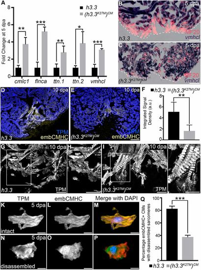
H3K27me3 deposition is required for sarcomere disassembly in wound edge cardiomyocytes. (A) Bar graph showing relative levels of sarcomeric transcripts in Tg(hsp70l:h3.3) and Tg(hsp70l:h3.3K27M)CM wound edge samples at 5 dpa, as measured by quantitative PCR (n=3 biological replicates for each cohort). Data are mean±s.d. *P<0.05; **P<0.01; ***P<0.001. (B,C) Section in situ hybridization for ventricular myosin (vmhcl) expression (blue) and antibody staining for myosin heavy chain (red). Control Tg(hsp70l:h3.3) hearts (B; n=6/6), but not Tg(hsp70l:h3.3K27M)CM hearts (C; n=5/5), exhibit reduced vmhcl expression in wound edge myocardium at 5 dpa. (D,E) Representative Tg(hsp70l:h3.3) (D) or Tg(hsp70l:h3.3K27M)CM (E) cardiac sections co-immunostained for tropomyosin (TPM; blue) and embCMHC (yellow). Tg(hsp70l:h3.3K27M)CM hearts showed relatively lower embCMHC signals at the wound edge compared with Tg(hsp70l:h3.3) hearts (n=8/8 and 11/11 hearts, respectively). (F) Bar graph showing the integrated signal densities of embCMHC in arbitrary units in Tg(hsp70l:h3.3) and Tg(hsp70l:h3.3K27M)CM hearts at 10 dpa. (G-J) Wound edge regions in cardiac sections immunostained for tropomyosin (TPM) at 10 dpa in Tg(hsp70l:h3.3) or Tg(hsp70l:h3.3K27M)CM hearts. Boxed regions in G and I are shown in H and J, respectively. Although wound edge cardiomyocytes show dissociated sarcomeres in Tg(hsp70l:h3.3) animals (n=5/5), sarcomere structure appears preserved in Tg(hsp70l:h3.3K27M)CM animals (n=4/6). (K-P) Split channel (K,L,N,O) or merged (M,P) confocal scans of dissociated cardiomyocytes immunostained for tropomyosin (TPM; red) and embryonic cardiac myosin heavy chain (embCMHC; green), and counterstained with DAPI (blue) to visualize sarcomere integrity in injury-responsive cardiomyocytes. (Q) The percentage of embCMHC+ cardiomyocytes with partially disassembled sarcomeres as shown in N-P in Tg(hsp70l:h3.3) (n=57 embCMHC+ CMs derived from nine apices) and Tg(hsp70l:h3.3K27M)CM (n=77 embCMHC+ CMs derived from nine apices) hearts. Data are mean±s.d. *P<0.05; **P<0.01; ***P<0.001. P-values were calculated using unpaired two-tailed Student's t-test. Scale bars: 100 µm in B-E; 10 µm in G-P.


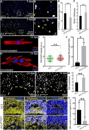
  • Injury-induced H3K27me3 deposition is required for cardiomyocyte cytokinesis and wound invasion during zebrafish heart regeneration. (A-D) Representative cardiac sections from heat-shocked Tg(hsp70l:h3.3) and Tg(hsp70l:h3.3K27M)CM animals at 5 dpa. Sections were double immunostained to identify cardiomyocyte nuclei (Mef2+; blue) and nuclei undergoing DNA replication (PCNA+; yellow). Boxed regions in A and C are shown at higher magnification in B and D, respectively. (E) The percentages of myocardial nuclei undergoing DNA replication near the wound edge were quantified and reported as mean proliferation indices (n=5 hearts for each cohort). Data are mean±s.d. n.s., not significant. (F) The percentages of myocardial nuclei that incorporated BrdU between 5 and 14 dpa near the wound edge were quantified and reported as mean proliferation indices [n=8 Tg(hsp70l:h3.3)n=5 Tg(hsp70l:h3.3K27M)CM]. (G,H) Fluorescent images of cardiomyocytes immunostained for tropomyosin (TPM) and counterstained with DAPI from dissociated Tg(hsp70l:h3.3) or Tg(hsp70l:h3.3K27M)CM ventricles following heat shock at 5 dpa. (I) The distribution of DNA content per nucleus in dissociated Tg(hsp70l:h3.3) (n=110 cells from 12 hearts) or Tg(hsp70l:h3.3K27M)CM (n=127 cells from 12 hearts) cardiomyocytes (as shown in G,H) based on DAPI fluorescence intensity. (J) The percentage of binucleated cardiomyocytes in Tg(hsp70l:h3.3) or Tg(hsp70l:h3.3K27M)CM ventricular dissociations. (K,L) Representative cardiac sections from heat-shocked Tg(hsp70l:h3.3) (n=3 hearts) and Tg(hsp70l:h3.3K27M)CM (n=5 hearts) animals at 14 dpa. Sections were immunostained to identify cardiomyocyte nuclei (Mef2). (M) Graph quantifying the wound edge CM nuclear density (CM nuclei per 200 μm). (N-S) Representative cardiac sections from heat-shocked Tg(hsp70l:h3.3) (n=10 hearts) and Tg(hsp70l:h3.3K27M)CM (n=5 hearts) animals at 10 dpa immunostained for tropomyosin (TPM, blue) and counterstained with DAPI to highlight nuclei and phalloidin to visualize the actin cytoskeleton in cells localized within the wound region and spared area. (T) Quantification of CM nuclei within the wound region in heat-shocked Tg(hsp70l:h3.3) and Tg(hsp70l:h3.3K27M)CM hearts. Data are mean±s.d. *P<0.05; **P<0.01; n.s., not significant. P-values were calculated using unpaired two-tailed Student's t-test. Scale bars: 50 µm in A,C,K,L,N-S; 10 µm in B,D,G,H.


Acknowledgments
This image is the copyrighted work of the attributed author or publisher, and ZFIN has permission only to display this image to its users. Additional permissions should be obtained from the applicable author or publisher of the image. Full text @ Development