
|
Fig. 4
-
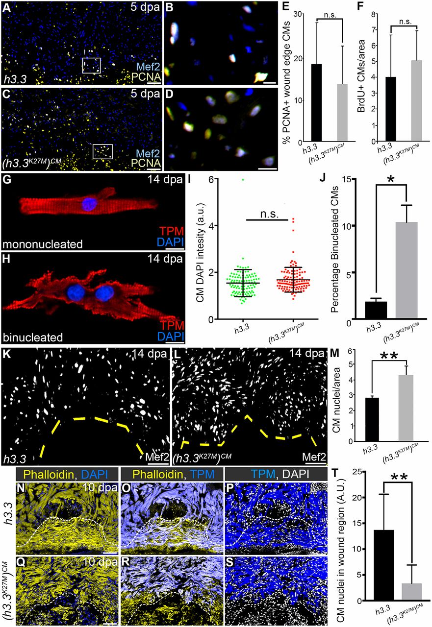
Injury-induced H3K27me3 deposition is required for cardiomyocyte cytokinesis and wound invasion during zebrafish heart regeneration. (A-D) Representative cardiac sections from heat-shocked Tg(hsp70l:h3.3) and Tg(hsp70l:h3.3K27M)CM animals at 5 dpa. Sections were double immunostained to identify cardiomyocyte nuclei (Mef2+; blue) and nuclei undergoing DNA replication (PCNA+; yellow). Boxed regions in A and C are shown at higher magnification in B and D, respectively. (E) The percentages of myocardial nuclei undergoing DNA replication near the wound edge were quantified and reported as mean proliferation indices (n=5 hearts for each cohort). Data are mean±s.d. n.s., not significant. (F) The percentages of myocardial nuclei that incorporated BrdU between 5 and 14 dpa near the wound edge were quantified and reported as mean proliferation indices [n=8 Tg(hsp70l:h3.3); n=5 Tg(hsp70l:h3.3K27M)CM]. (G,H) Fluorescent images of cardiomyocytes immunostained for tropomyosin (TPM) and counterstained with DAPI from dissociated Tg(hsp70l:h3.3) or Tg(hsp70l:h3.3K27M)CM ventricles following heat shock at 5 dpa. (I) The distribution of DNA content per nucleus in dissociated Tg(hsp70l:h3.3) (n=110 cells from 12 hearts) or Tg(hsp70l:h3.3K27M)CM (n=127 cells from 12 hearts) cardiomyocytes (as shown in G,H) based on DAPI fluorescence intensity. (J) The percentage of binucleated cardiomyocytes in Tg(hsp70l:h3.3) or Tg(hsp70l:h3.3K27M)CM ventricular dissociations. (K,L) Representative cardiac sections from heat-shocked Tg(hsp70l:h3.3) (n=3 hearts) and Tg(hsp70l:h3.3K27M)CM (n=5 hearts) animals at 14 dpa. Sections were immunostained to identify cardiomyocyte nuclei (Mef2). (M) Graph quantifying the wound edge CM nuclear density (CM nuclei per 200 μm). (N-S) Representative cardiac sections from heat-shocked Tg(hsp70l:h3.3) (n=10 hearts) and Tg(hsp70l:h3.3K27M)CM (n=5 hearts) animals at 10 dpa immunostained for tropomyosin (TPM, blue) and counterstained with DAPI to highlight nuclei and phalloidin to visualize the actin cytoskeleton in cells localized within the wound region and spared area. (T) Quantification of CM nuclei within the wound region in heat-shocked Tg(hsp70l:h3.3) and Tg(hsp70l:h3.3K27M)CM hearts. Data are mean±s.d. *P<0.05; **P<0.01; n.s., not significant. P-values were calculated using unpaired two-tailed Student's t-test. Scale bars: 50 µm in A,C,K,L,N-S; 10 µm in B,D,G,H.