
- Title
-
The role of internal convection in respiratory gas transfer and aerobic metabolism in larval zebrafish ( Danio rerio)
- Authors
- Hughes, M.C., Zimmer, A.M., Perry, S.F.
- Source
- Full text @ Am. J. Physiol. Regul. Integr. Comp. Physiol.
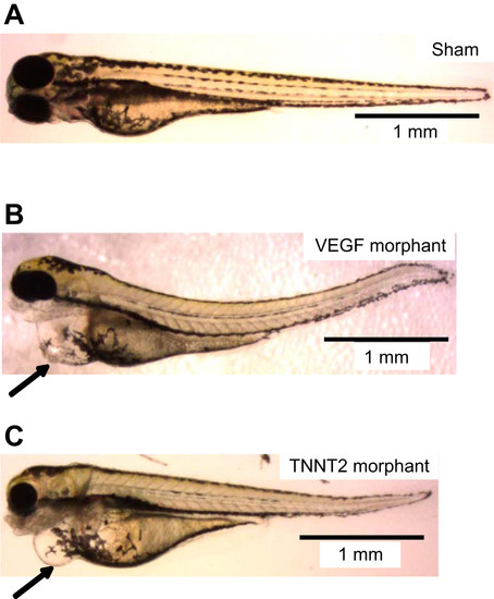
Representative microscopy images of sham (A), VEGF morphant (B), and cardiac troponin T (TNNT2; C) morphant zebrafish larvae at 4 days postfertilization (dpf). Black arrows indicate pericardial edema; scale bars represent 1 mm.
|
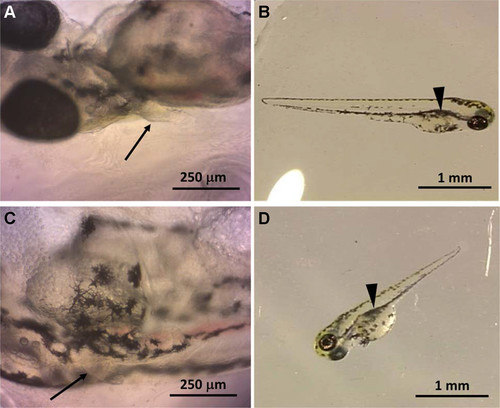
Representative microscopy images of sham (A and B) and VEGF morphant (C and D) larvae at 60 h postfertilization (hpf; A and C) and 72 hpf (B and D). Arrows indicate pectoral fins, and arrowheads indicate melanin accumulation overlying the swimbladder rudiment.
|
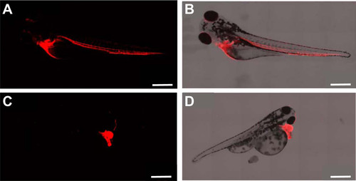
Whole body microscopic images of 4 dpf sham larvae (A and B) and VEGF morphants (C and D) injected with fluorescently tagged microspheres. A and C: fluorescence images. B and D: fluorescence and transmitted light overlays. Scale bars represent 500 µm.
|