
- Title
-
Evolution of Endothelin signaling and diversification of adult pigment pattern in Danio fishes
- Authors
- Spiewak, J.E., Bain, E.J., Liu, J., Kou, K., Sturiale, S.L., Patterson, L.B., Diba, P., Eisen, J.S., Braasch, I., Ganz, J., Parichy, D.M.
- Source
- Full text @ PLoS Genet.

Phylogenetic relationship and pigment patterns of D. rerio and D. nigrofasciatus. (A) Phylogenetic relationships of selected Danio species [9]. (B) Danio rerio exhibit several dark stripes of melanophores with sparse iridophores, and light interstripes with abundant iridophores. Danio nigrofasciatus share common pattern elements but have fewer stripes and interstripes overall with spots forming ventrally instead of stripes. A shiny ventrum in both species results principally from iridophores that line the peritoneum, rather than iridophores in the hypodermis of the skin. Insets show iridescence of hypodermal iridophores. |

Iridophore development differs between D. rerio and D. nigrofasciatus. (A) Young adult patterns of the two species, illustrating fewer melanophores of D. nigrofasciatus compared to D. rerio. Stripes and interstripes are marked at the left. 1D, 1V: primary dorsal and ventral stripes. 2V, secondary ventral stripe. I1, I2, I3: Primary, secondary and tertiary interstripes. (B) Iridophores during primary stripe and secondary interstripe formation. Shown are representative individuals imaged repeatedly for D. rerio (upper) and D. nigrofasciatus (lower), with iridophores of the primary interstripe indicated by blue arrowheads. Fish were imaged throughout adult pattern formation with stages PB through J [14] illustrated here (corresponding to days ~14 through 38 post fertilization, shown for heuristic purposes only). Insets show iridophores at higher magnification. In D. nigrofasciatus, iridophores are comparatively few, and do not as extensively populate the region of the secondary ventral stripe or the secondary ventral interstripe (blue arrow in D. rerio). Melanophores of the primary ventral stripe occur more ventrally than in D. rerio and tended to be more closely associated with vertical myosepta (marked by red asterisks). Apparent differences in melanophore sizes between species may reflect lower densities and more spread morphologies in D. nigrofasciatus compared to D. rerio [26,79]. Sample sizes (N): 9 D. rerio; 6 D. nigrofasciatus. (C) Clonally related iridophores increased in number in both species between formation of primary interstripe (left; stage PB+) and subsequent pattern reiteration (right; J++). Points connected by lines represent individuals at each developmental stage. Starting numbers were not significantly different (F1,10 = 0.94, P = 0.4), whereas final numbers were significantly fewer in D. nigrofasciatus than in D. rerio (repeated measures, species x stage interaction, F1,10 = 7.47, P<0.05; N = 5 D. rerio, N = 6 D. nigrofasciatus). |

Shown are wild-type (wt) and homozygous mutants for edn3a and edn3b, double mutant edn3a; edn3b, and ednrb1a. edn3a mutants had normal hypodermal pigment pattern, including iridophore interstripes (arrow, right) but lacked peritoneal iridophores (arrowhead). edn3b mutants had hypodermal iridophore and melanophore deficiencies but normal peritoneal iridophores. Fish doubly mutant for these loci exhibited both defects and resembled ednrb1a mutants. At the very young adult stages shown, ventral-most melanophores of ednrb1a mutants (*) have yet to coalesce into spots. |

ZFIN is incorporating published figure images and captions as part of an ongoing project. Figures from some publications have not yet been curated, or are not available for display because of copyright restrictions. PHENOTYPE:
|
|
ZFIN is incorporating published figure images and captions as part of an ongoing project. Figures from some publications have not yet been curated, or are not available for display because of copyright restrictions. PHENOTYPE:
|
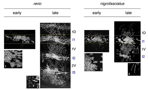
Expansion of iridophore clones differs between D. rerio and D. nigrofasciatus. Representative images for individuals of each species mosaic for iridophore reporter pnp4a:palmEGFP at an early stage of pattern formation, and a late stage, once patterns were complete. Dashed yellow lines indicate approximate regions of correspondence between early and late images and I1–I3 indicate primary through tertiary interstripes, if present; 1D, 1V, 2V indicate positions of stripes, if present. In each species, iridophores were present within interstripes, where they were densely packed, and within stripe, where they were loosely arranged. Inset 1, clonal derived early iridophores in primary interstripe of D. rerio. Inset 2, In some individuals, autofluorescent xanthophores (x) were apparent but were distinguishable from iridophores by differences in shape. Inset 3, early iridophores of D. nigrofasciatus. Inset 4, Examples of spindle-shaped “type-L” iridophores [79] present at low abundance in each species. |