
Fig. S1
- ID
- ZDB-FIG-181108-8
- Publication
- Spiewak et al., 2018 - Evolution of Endothelin signaling and diversification of adult pigment pattern in Danio fishes
- Other Figures
- All Figure Page
- Back to All Figure Page
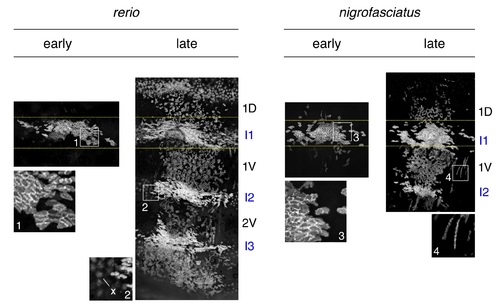
Expansion of iridophore clones differs between D. rerio and D. nigrofasciatus. Representative images for individuals of each species mosaic for iridophore reporter pnp4a:palmEGFP at an early stage of pattern formation, and a late stage, once patterns were complete. Dashed yellow lines indicate approximate regions of correspondence between early and late images and I1–I3 indicate primary through tertiary interstripes, if present; 1D, 1V, 2V indicate positions of stripes, if present. In each species, iridophores were present within interstripes, where they were densely packed, and within stripe, where they were loosely arranged. Inset 1, clonal derived early iridophores in primary interstripe of D. rerio. Inset 2, In some individuals, autofluorescent xanthophores (x) were apparent but were distinguishable from iridophores by differences in shape. Inset 3, early iridophores of D. nigrofasciatus. Inset 4, Examples of spindle-shaped “type-L” iridophores [79] present at low abundance in each species. |