FIGURE SUMMARY
Title

Development of brain ventricular system

Authors
Korzh, V.
Source
Full text @ Cell. Mol. Life Sci.

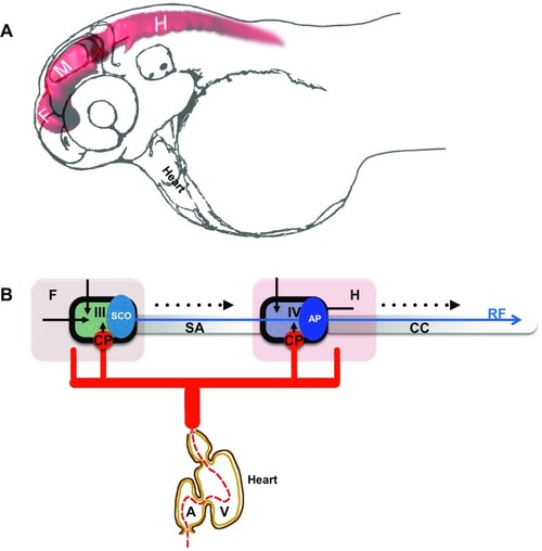
Development of the brain ventricular system of zebrafish. a The dye-filled ventricular system of 48 hpf zebrafish consists of three distinct cavities, which are reshaped as two ventricles and sylvius aqueduct connecting these. b Hypothetical scheme connecting the zebrafish blood circulation and the brain ventricular systems. During development influx of eCSF inflates and shapes the brain ventricular system [50, 51]. The choroid plexi contribute CSF enriched by ions, micronutrients and proteins. The SCO produces substances of the Reissner fiber, etc. A atrium, AP area postrema, CC central canal, CP choroid plexus, F forebrain, H hindbrain, M midbrain, SA Sylvius aqueduct, SCO subcommissural organ, V heart ventricle, III III brain ventricle, IV IV brain ventricle. Black arrow indicates embryonic CSF influx, arrowhead indicates CSF influx, broken line indicates direction of CSF flow. Blue arrow indicates Reissner fiber, red solid lines indicates vasculature and black solid line indicates ependyma

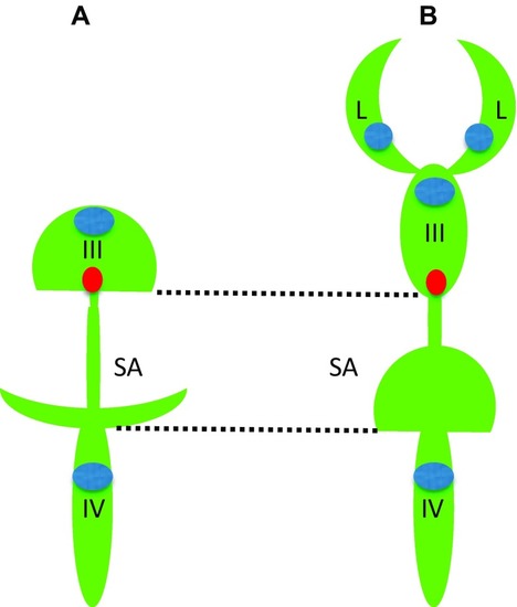
Comparison of brain ventricular system in 6 dpf zebrafish (a) and stage 53 Xenopus (b). The zebrafish larval BVS contains two ventricles unlike that of larval Xenopus. Whereas the IIIrd ventricle and posterior SA by the end of embryogenesis acquire rather complex configuration these are not that different from corresponding part of BVS in Xenopus. Green—ventricular system, blue—choroid plexus, red—subcommissural organ, III—IIIrd ventricle, IV—IVth ventricle, L—lateral ventricle, SA—Sylvius aqueduct

(modified from Turner et al. [77], and Mogi et al. [58], correspondingly)

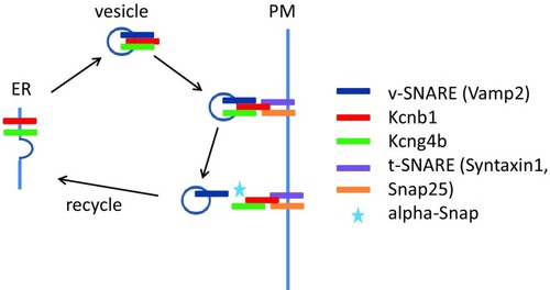
Genetic analyses reveal a role of the Kcnb1-Kcng4 axis in development of the BVS. Mutation/LOF of Kcg4b causes strong hydrocephalus and GOF—almost complete failure of ventricle inflation. In contrast, Kcnb1 mutant shows the slit-ventricle phenotype. The defect of Kcnb1 not only eliminates function of this protein as an electrically active subunit of voltage-gated Kv channel. Given its role in transport of Kcng4b, the defect of Kcnb1 negatively impacts function of both proteins, which counteract each other activity at the plasma membrane. Thus, the complete LOF of Kcnb1 could be partially compensated by a deficiency of Kcng4b transport. Note that Kcng4b is not expressed in adult animals suggesting that its modulation of Kcnb1 activity takes place only during development

Acknowledgments
This image is the copyrighted work of the attributed author or publisher, and ZFIN has permission only to display this image to its users. Additional permissions should be obtained from the applicable author or publisher of the image. Full text @ Cell. Mol. Life Sci.