
- Title
-
Mapping a multiplexed zoo of mRNA expression
- Authors
- Choi, H.M., Calvert, C.R., Husain, N., Huss, D., Barsi, J.C., Deverman, B.E., Hunter, R.C., Kato, M., Lee, S.M., Abelin, A.C., Rosenthal, A.Z., Akbari, O.S., Li, Y., Hay, B.A., Sternberg, P.W., Patterson, P.H., Davidson, E.H., Mazmanian, S.K., Prober, D.A., van de Rijn, M., Leadbetter, J.R., Newman, D.K., Readhead, C., Bronner, M.E., Wold, B., Lansford, R., Sauka-Spengler, T., Fraser, S.E., Pierce, N.A.
- Source
- Full text @ Development
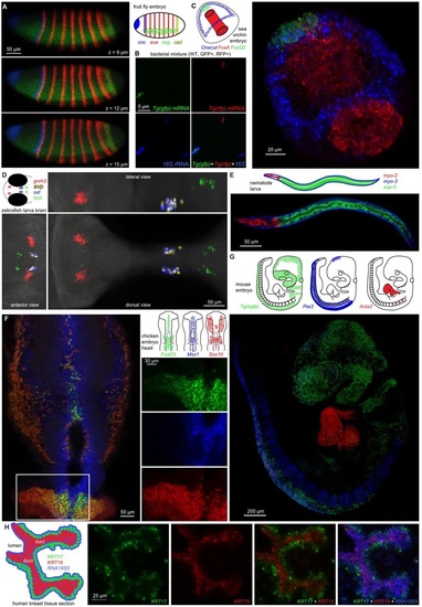
Multiplexed mRNA expression maps using in situ HCR. (A) Whole-mount fruit fly (Drosophila melanogaster) embryo: expression schematic and confocal micrographs for four target mRNAs on three planes. Embryo fixed: stage 4-6. (B) Mixed bacterial populations (Escherichia coli: WT, GFP+, RFP+): epifluorescence micrographs (single channels and merge) for three targets (gfp and rfp mRNAs and 16S rRNA). (C) Whole-mount sea urchin embryo (Strongylocentrotus purpuratus): expression schematic and three-dimensional reconstruction from confocal micrographs for three target mRNAs. Embryo fixed: 45 hpf. (D) Whole-mount zebrafish larva (Danio rerio): expression schematic and three-dimensional reconstruction from confocal micrographs for four target mRNAs within the brain. Larva fixed: 5 dpf. (E) Whole-mount nematode larva (Caenorhabditis elegans): expression schematic and confocal micrograph for three target mRNAs. Larva fixed: L3. (F) Whole-mount chicken embryo (Gallus gallus domesticus): expression schematic and confocal micrographs for three target mRNAs in the neural crest (merge and single-channel details). Embryo fixed: stage HH 11-12. (G) Whole-mount mouse embryo [Mus musculus: Tg(Wnt1-Cre; R26R-eGFP)]: expression schematic and three-dimensional reconstruction from confocal micrographs for three target mRNAs. Embryo fixed: E9.5. (H) FFPE human breast tissue section (Homo sapiens sapiens): expression schematic and epifluorescence micrographs for two target mRNAs and one rRNA (single channels and merges). Thickness: 4 µm. See Figs S2-S10 and Movies 1-5 for additional data. |
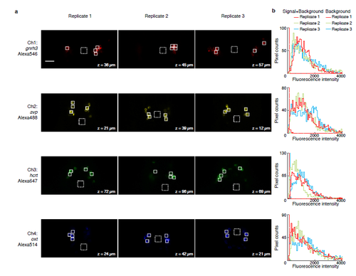
Multiplexed in situ HCR in whole-mount zebrafish larvae (D. rerio). (a) Individual channels from 4-channel confocal images. For each of three replicate larvae, a representative optical section was selected for each channel based on the expression depth of the corresponding target mRNA. (b) Pixel intensity histograms for Signal + Background (pixels within solid boundary) and Background (pixels within dashed boundary). For each image, the total number of pixels within solid and dashed boundaries is the same. Larvae fixed: 5 dpf. Scale bar: 25 μm. |