
Image
|
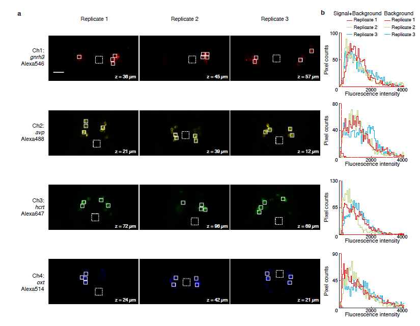
Figure Caption
Fig. S6
Multiplexed in situ HCR in whole-mount zebrafish larvae (D. rerio). (a) Individual channels from 4-channel confocal images. For each of three replicate larvae, a representative optical section was selected for each channel based on the expression depth of the corresponding target mRNA. (b) Pixel intensity histograms for Signal + Background (pixels within solid boundary) and Background (pixels within dashed boundary). For each image, the total number of pixels within solid and dashed boundaries is the same. Larvae fixed: 5 dpf. Scale bar: 25 μm.
Acknowledgments
This image is the copyrighted work of the attributed author or publisher, and
ZFIN has permission only to display this image to its users.
Additional permissions should be obtained from the applicable author or publisher of the image.
Full text @ Development